| CPC A61B 8/0825 (2013.01) [A61B 8/085 (2013.01); A61B 8/483 (2013.01); G06T 7/11 (2017.01); G06T 7/149 (2017.01); G06T 17/00 (2013.01); G06V 10/143 (2022.01); G06V 10/25 (2022.01); G06V 10/50 (2022.01); G06V 10/75 (2022.01); G06T 2207/10132 (2013.01); G06T 2207/20081 (2013.01); G06T 2207/30068 (2013.01)] | 20 Claims |

|
1. An analysis method for breast ultrasound images, comprising:
obtaining at least one breast ultrasound image, wherein the at least one breast ultrasound image is used for forming a three dimensional (3D) breast model;
obtaining a volume of interest (VOI) as a suspicious area in the at least one breast ultrasound image by applying a detection model on the 3D breast model, wherein the detection model is trained by a machine learning algorithm;
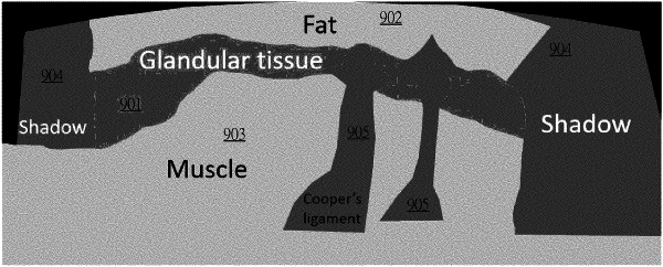
segmenting the at least one breast ultrasound image into a plurality of tissues, to obtain a tissue segmentation result, wherein the tissue segmentation result comprises the plurality of tissues in the 3D breast model;
determining a type of a target tissue of the plurality of tissue where the VOI is located with the tissue segmentation result;
determining the VOI as a false positive according to the target tissue where the VOI is located, wherein the target tissue where the VOI is located is a glandular tissue based on the tissue segmentation result, and determining the VOI as the false positive according to the target comprises:
determining whether the VOI located in the glandular tissue is further located in a lactiferous duct based on the tissue segmentation result;
in response to the VOI being located in the glandular tissue but not located in the lactiferous duct, determining the VOI as not the false positive; and
in response to the VOI being located in both the glandular tissue and the lactiferous duct, determining the VOI as the false positive; and
removing the VOI belonging to the false positive from the suspicious area.
|