| CPC G06V 20/695 (2022.01) [G06V 10/235 (2022.01); G06V 10/993 (2022.01)] | 18 Claims |
|
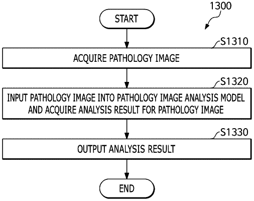
1. A method for analysing a pathology image, the method being performed by at least one processor and comprising:
acquiring a pathology image;
generating an analysis result for the pathology image based on the pathology image by using a pathology image analysis model configured to:
generate a first analysis result by analysing a staining intensity at a first location where a staining color is expressed on at least one cell;
generate a second analysis result by analysing a staining intensity at a second location where the staining color is expressed on the at least one cell;
classify a first cell and a second cell in the pathology image based on staining intensity amounts at the first location on the first cell and the first location on the second cell in the pathology image;
segment the pathology image into a first segment corresponding to the first cell and a second segment corresponding to the second cell; and
visualize the first segment in a first color, and the second segment in a second color different from the first color; and
outputting the analysis result including at least one of the first analysis result or the second analysis result,
wherein the first location and the second location include at least one of cell membrane, cell nucleus, or cytoplasm.
|