| CPC A61B 5/0059 (2013.01) [A61B 5/0033 (2013.01); A61B 5/0035 (2013.01); A61B 5/0036 (2018.08); A61B 5/0037 (2013.01); A61B 5/0066 (2013.01); A61B 5/0073 (2013.01); A61B 5/0079 (2013.01); A61B 5/0088 (2013.01); A61B 5/4542 (2013.01); A61B 5/4547 (2013.01); A61B 5/682 (2013.01); A61B 5/748 (2013.01); A61B 5/7485 (2013.01); A61C 9/0053 (2013.01); A61C 9/006 (2013.01); A61C 19/04 (2013.01); A61B 5/0064 (2013.01); A61B 5/4848 (2013.01); A61B 5/6849 (2013.01); A61B 5/743 (2013.01)] | 5 Claims |

|
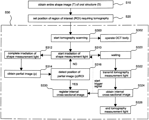
1. An intraoral tomography method comprising:
sequentially irradiating pieces of shape measurement light onto an oral structure, detecting reflected light formed when the shape measurement light is reflected from a surface of the oral structure, and obtaining an entire image (T) of the oral structure;
setting a position of a region of interest (ROI) requiring tomography for the entire image (T) of the oral structure;
transmitting tomography measurement light along the set ROI, detecting reflected light reflected inside the ROI, and
obtaining an internal cross-sectional image of the ROI, wherein the internal cross-sectional image of the ROI is obtained by steps comprising:
irradiating the shape measurement light onto a part of the oral structure(S), detecting reflected light formed when the shape measurement light is reflected from a surface of the part of the oral structure(S), and obtaining a partial image (p) of the oral structure;
comparing the partial image (p) of the oral structure(S) with the entire image (T) of the oral structure(S), detecting a position of the partial image (p) in the entire image (T) of the oral structure(S), and detecting whether the ROI is present in the partial image (p);
transmitting the tomography measurement light to the part of the oral structure(S) onto which the shape measurement light has been irradiated, detecting reflected light reflected inside the oral structure(S), and obtaining an internal cross-sectional image of the oral structure(S); and
when the ROI is present in the partial image (p), registering the internal cross-sectional image, which is obtained from the part of the oral structure(S) onto which the shape measurement light has been irradiated, as the internal cross-sectional image of the ROI of the partial image (p).
|