| CPC G06V 10/778 (2022.01) [G06T 7/11 (2017.01); G06T 7/136 (2017.01); G06T 7/194 (2017.01); G06V 10/764 (2022.01); G06V 10/776 (2022.01); G06V 10/86 (2022.01); G06V 20/50 (2022.01); G06V 20/70 (2022.01); G16H 30/40 (2018.01); G16H 70/60 (2018.01); G06T 2207/20021 (2013.01); G06T 2207/20081 (2013.01); G06T 2207/20084 (2013.01); G06T 2207/30024 (2013.01); G06T 2207/30061 (2013.01); G06T 2207/30068 (2013.01); G06T 2207/30096 (2013.01); G06V 2201/03 (2022.01)] | 7 Claims |

|
1. A weakly supervised pathological image tissue segmentation method based on an online noise suppression strategy, comprising:
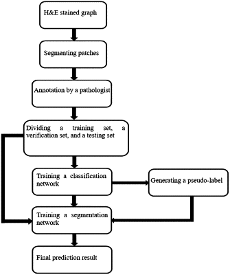
acquiring an H&E stained graph, processing the H&E stained graph to obtain a data set, dividing the data set, training a classification network based on a divided data set, and generating a pseudo-label;
suppressing a noise existing in the pseudo-label based on the online noise suppression strategy, and training a semantic segmentation network through the pseudo-label after noise suppression and a training set corresponding to the pseudo-label to obtain a prediction result of the semantic segmentation network after the training, and taking the prediction result as a final segmentation result;
acquiring the H&E stained graph comprises:
collecting pathological section images of cancerous tissues of lung cancer/breast cancer patients, dyeing the pathological section images to obtain H&E stained pathological sections of lung cancer/breast cancer, and then digitizing the H&E stained pathological sections of the lung cancer/breast cancer to obtain the H&E stained graph;
processing the H&E stained graph comprises:
delineating a region of interest of the H&E stained graph, cutting the region of interest into sub-image blocks of a same series without overlapping, adding a patch-level label to each of the sub-image blocks, and specifying a tissue category existing in the patch-level label;
dividing the data set, training the classification network based on the divided data set, and generating the pseudo-label comprise:
dividing the training set, a verification set and a test set according to the data set, constructing the classification network by using a deep learning model, carrying out data enhancement processing on the training set, training the classification network based on the training set after the data enhancement processing, internally verifying classification performance of the classification network through the verification set, and externally verifying the classification performance of the classification network through the test set to obtain a trained classification network, and using Grad-CAM++ to generate the pseudo-label based on the trained classification network.
|