| CPC G01R 33/281 (2013.01) [A61B 5/0042 (2013.01); A61B 5/055 (2013.01); A61B 5/4842 (2013.01); A61B 5/4848 (2013.01); A61B 5/7275 (2013.01); G01R 33/4806 (2013.01); G01R 33/5601 (2013.01); G01R 33/5608 (2013.01); G06T 7/0016 (2013.01); G16H 30/40 (2018.01); G16H 40/40 (2018.01); G16H 40/63 (2018.01); A61B 2576/026 (2013.01); A61K 31/166 (2013.01); A61K 31/555 (2013.01); A61K 31/7068 (2013.01); A61K 2300/00 (2013.01); G06T 2207/10096 (2013.01); G06T 2207/30016 (2013.01); G06T 2207/30096 (2013.01)] | 28 Claims |

|
1. Apparatus for operating MRI, comprising:
a control for operating an MRI scanner to carry out an MRI scan of an organ of a subject, the control being configured to carry out a first MRI scan at a beginning of a predetermined time interval, said beginning being at least three minutes and no more than twenty minutes post contrast administration, and a second MRI scan at an end of said predetermined time interval, said end being at least twenty minutes after said beginning of said predetermined time interval;
an input for receiving said first and said second MRI scans;
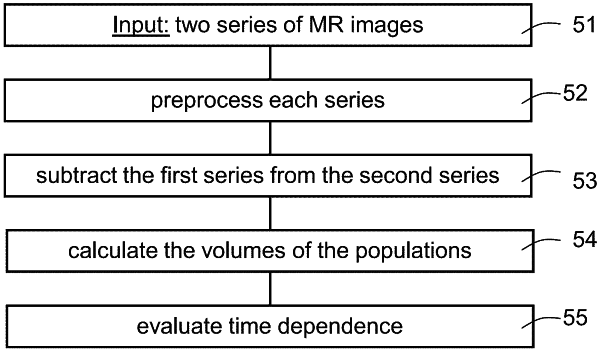
an image processor having a circuit configured for forming a subtraction map from said first and said second MRI scans by analyzing said scans to distinguish between two primary populations, a slow population, in which contrast clearance from the tissue is slower than contrast accumulation, and a fast population in which clearance is faster than accumulation; and
an output converting said scans to a displayed subtraction map on which distributions of said two primary populations are distinguished, wherein a distribution of said fast population corresponds to a tumoral tissue region and a distribution of said slow population corresponds to a non-tumoral tissue region;
wherein said control is configured to carry out said first scan at said beginning of said predetermined time interval, and to carry out said second scan at said end of said predetermined time interval;
wherein said image processor is configured for carrying out registration between corresponding MRI scans; and
wherein the apparatus is in use for differentiating tumor from treatment effects.
|