| CPC A61B 90/36 (2016.02) [A61B 8/0841 (2013.01); A61B 8/12 (2013.01); A61B 10/0045 (2013.01); A61B 18/1477 (2013.01); A61M 5/46 (2013.01); A61B 2010/045 (2013.01); A61B 2018/1425 (2013.01); A61B 2034/107 (2016.02); A61B 90/11 (2016.02); A61B 2090/378 (2016.02)] | 27 Claims |

|
1. A system for treating uterine anatomy, the system comprising:
a radiofrequency ablation device comprising a needle array comprising multiple needles, wherein the needle array is deployable into a uterine fibroid;
a radiofrequency generator; and
a processing unit programmed to:
display, on a screen, a real-time image of the uterine anatomy including a uterine fibroid to be treated;
overlay, onto the real-time image, a projected path of the radiofrequency ablation device;
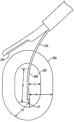
overlay, onto the real-time image, a projected treatment boundary based on a first, actual position of the needle array prior to deployment of the needle array into the uterine fibroid;
recalculate the projected treatment boundary based on a second, actual position of the needle array after deployment of the needle array into the uterine fibroid;
overlay, onto the real-time image, the recalculated projected treatment boundary; and
deliver radiofrequency energy from the radiofrequency generator through the needle array to treat the uterine fibroid after recalculating the projected treatment boundary and with at least a portion of the uterine fibroid positioned within the recalculated projected treatment boundary on the real-time image.
|