| CPC A61B 5/435 (2013.01) [A61B 1/00172 (2013.01); A61B 1/07 (2013.01); A61B 1/303 (2013.01); A61B 5/0095 (2013.01); A61B 5/02007 (2013.01); G02B 26/0833 (2013.01); G02B 26/105 (2013.01); G02F 1/17 (2013.01); A61B 2562/028 (2013.01); G02F 2203/48 (2013.01)] | 5 Claims |

|
1. A method of imaging a microvasculature within a tissue using a fast-scanning optical-resolution photoacoustic (fsOR-PAE) system, the method comprising:
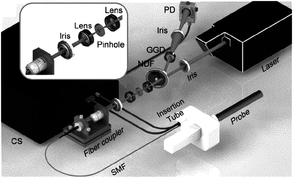
providing an fsOR-PAE endoscope comprising a scanning mirror and an ultrasonic transducer housed within an elongate housing, a set of doublets positioned between a pulsed laser source and the scanning mirror, and the pulsed laser source, the pulsed laser source and the ultrasonic transducer coupled to the scanning mirror;
positioning the elongate housing within the tissue;
producing a plurality of laser pulses using the pulsed laser;
focusing the plurality of laser pulses using the set of doublets pulses;
directing the plurality of laser pulses through an imaging window formed within the housing to a plurality of focal spots within the tissue in a scanning pattern using the scanning mirror, wherein directing the plurality of laser pulses in a scanning pattern comprises scanning in a sweep parallel to cylindrical axis and a B-scan rate of 250 Hz over a 3 mm range;
receiving and directing a plurality of photoacoustic signals produced at the plurality of focal spots to the ultrasonic transducer via the imaging window;
detecting the plurality of photoacoustic signals using the ultrasonic transducer; and
transforming the plurality of detected photoacoustic signals into an image of the microvasculature, wherein the image comprises a lateral resolution of less than 5 μm.
|