| CPC A61B 5/062 (2013.01) [A61B 1/267 (2013.01); A61B 5/113 (2013.01); A61B 5/7207 (2013.01); A61B 8/12 (2013.01); A61B 8/4254 (2013.01); A61B 8/5238 (2013.01); A61B 8/08 (2013.01); A61B 8/463 (2013.01); A61B 8/467 (2013.01); A61B 8/5261 (2013.01)] | 20 Claims |
|
1. A lung visualization system using ultrasound (US) imaging, the lung visualization system comprising:
a memory storing a first three dimensional (3D) model of a luminal network of a lung;
an electromagnetic (EM) board configured to generate an EM field;
a first catheter configured to navigate the luminal network toward a target;
a second catheter configured to be inserted through the first catheter;
an EM sensor configured to sense the EM field;
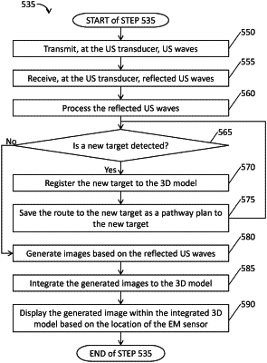
an US transducer configured to transmit US waves and generate US images based on the US waves reflected from the luminal network, the EM sensor positioned at a distal portion of one of the first and second catheters and the US transducer positioned at a distal portion of the other one of the first and second catheters;
a processor configured to:
process the sensed EM field to measure a travelling distance of the EM sensor;
scale the travelling distance so that the scaled distance is matched to a coordinate system of the first 3D model;
replace lower resolution portions of Computed Tomography (CT) image data from which the first 3D model was generated with the generated US images at a location of the US transducer based on the scaling of the travelling distance, yielding a modified CT image data;
generate a second 3D model from the modified CT image data, wherein the second 3D model has a higher resolution than the first 3D model; and
a display device configured to display the second 3D model.
|