| CPC G06T 7/0012 (2013.01) [G06T 11/006 (2013.01); G16H 30/40 (2018.01); G06T 2207/20081 (2013.01); G06T 2207/30016 (2013.01); G06T 2207/30096 (2013.01)] | 10 Claims |

|
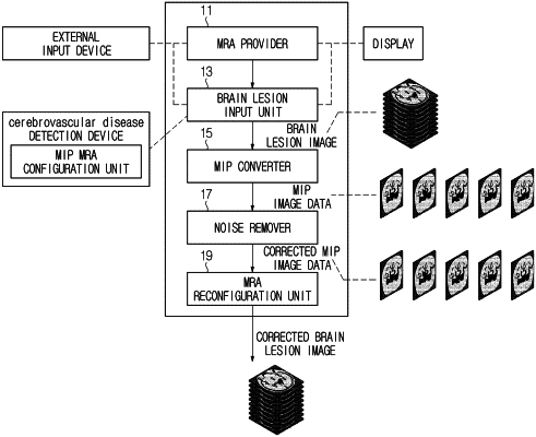
1. An electronic apparatus for providing brain lesion information based on an image, the electronic apparatus comprising:
a magnetic resonance angiography (MRA) provider configured to provide an environment capable of displaying 3D time-of-flight magnetic resonance angiography (3D TOF MRA) using user input;
a brain lesion input unit configured to annotate a brain lesion area and to generate and manage a brain lesion image by combining the annotated brain lesion area and the 3D TOF MRA;
a maximum intensity projection (MIP) converter configured to configure MIP image data including at least one image frame corresponding to a projection position of the brain lesion image;
a noise remover configured to configure at least one three-dimensional sinogram using the brain lesion image, remove noise of the at least one three-dimensional sinogram, and configure corrected MIP image data reflecting brain lesion information based on the noise-removed three-dimensional sinogram; and
an MRA reconfiguration unit configured to reconfigure a corrected brain lesion image by back-projecting the corrected MIP image data.
|