| CPC G06T 5/005 (2013.01) [G06F 18/24 (2023.01); G06T 5/001 (2013.01); G06T 5/50 (2013.01); G06T 7/11 (2017.01); G06T 7/174 (2017.01); G06T 11/008 (2013.01); G06T 2207/10088 (2013.01); G06T 2207/10104 (2013.01); G06T 2207/20152 (2013.01); G06T 2207/20224 (2013.01); G06T 2211/432 (2013.01); G06V 2201/03 (2022.01)] | 13 Claims |

|
1. A truncation cotnpensation system, comprising:
a PET image memory configured to store a volume of PET imaging data for a PET imaging volume;
a MR image memory configured to store a volume of MR imaging data having a MR field of vision (FOV), wherein the MR FOV is smaller than the PET imaging volume;
one or more processors configured to:
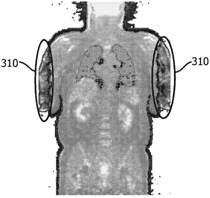
mask the volume of the PET imaging data with the volume of the MR imaging data to generate a masked PET image that includes one or more truncated regions of interest (ROIs) that are outside of the MR FOV;
identify the one or more truncated ROIs in the masked PET image from the masking of the volume of the PET imaging data with the volume of the MR imaging data;
classify the one or more truncated ROIs as types of anatomical structures that are outside the MR FOV based on structure and characteristics for each truncated ROI, the structure of each truncated ROI including a thickness, shape, and size of the corresponding truncated ROI;
classify each classified anatomical structure as to whether it is inside of or outside of a MR FOV of the PET imaging data;
compensate for truncation in the truncated ROIs based on the classified anatomical structure in the masked PET image that is outside the MR FOV to generate compensated ROIs; and
merge the compensated ROIs with the MR imaging data to form a compensated MR map.
|