| CPC A61B 34/20 (2016.02) [A61B 8/0841 (2013.01); A61B 8/4488 (2013.01); A61B 8/463 (2013.01); A61B 2034/2063 (2016.02)] | 2 Claims |

|
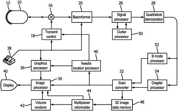
1. A method for operating an ultrasonic imaging system for image guidance of needle insertion in a subject, the method comprising:
acquiring a first ultrasound image of an image field in the subject with a curved array transducer probe, the image field suspected of comprising a needle inserted into the subject, the ultrasound image comprising a plurality of echo returns from unsteered transmit beams impinging upon the needle at a plurality of different angles of incidence;
identifying an angle of a transmit beam for producing a peak magnitude of echo return from the needle in the image field based on application of image processing to said ultrasound image echo returns;
transmitting a plurality of parallel steered beams from the curved array transducer probe at the identified angle and receiving image data in response;
processing the image data with two different apodization functions adapted to isolate clutter in the image data and forming two images, each using a different apodization function;
correlating or combining pixel values of the two images processed with the two different apodization functions to produce clutter-reduced image data; and
displaying a clutter-reduced ultrasound image of the needle in the image field;
wherein processing the image data with two different apodization functions comprises processing the image data with complementary apodization functions,
wherein processing the image data with complementary apodization functions comprises processing the image data with apodization functions which affect side lobe artifact data differently, and
wherein using image data processed with the two different apodization functions comprises combining image data having both side lobe and main lobe data with image data having only side lobe or main lobe data.
|