| CPC G06T 7/0012 (2013.01) [G06F 16/538 (2019.01); G06T 7/13 (2017.01); G06T 7/136 (2017.01); G06T 7/60 (2013.01); G16H 30/40 (2018.01); G06T 2207/10081 (2013.01); G06T 2207/10088 (2013.01); G06T 2207/20044 (2013.01); G06T 2207/30008 (2013.01)] | 11 Claims |
|
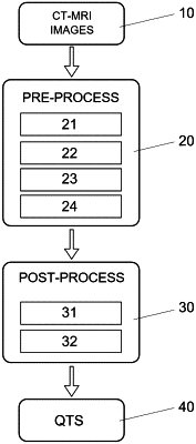
1. A method for obtaining an image biomarker that quantifies the quality of the trabecular structure of bones, the method comprising:
retrieving high-resolution trabecular images generated by a technique selected from among Computed Tomography (CT), Magnetic Resonance Imaging (MRI) and a combination of CT and MRI, wherein the high-resolution trabecular images are retrieved from a medical image database with a large quantity of content from trabecular regions;
pre-processing the high-resolution trabecular images, wherein pre-processing the high-resolution trabecular images comprises:
obtaining a region of interest (ROI);
calculating a bone fraction map;
removing a partial volume effect; and
binarizing;
post-processing the high-resolution trabecular images, wherein post-processing the high-resolution trabecular images comprises:
skeletonisation; and
extracting morphological and structural characteristics; and
obtaining a unique image biomarker (QTS) based on the following equation:
QTS=0.7137*Comp1+0.2863*Comp2,
where:
Comp1=BV/TV1*BV/TV+TbTh1*TbTh+TbSp1*TbSp+TbN1*TbN+D2D1*D2D+D3D1*D3D;
Comp2=BV/TV2*BV/TV+TbTh2*TbTh+TbSp2*TbSp+TbN2*TbN+D2D2*D2D+D3D2*D3D,
where:
BV/TV1=0.255; TbTh1=−0.023; TbSp1=−0.277; TbN1=0.280; D2D1=0.246; D3D1=0.089;
BV/TV2=0.331; TbTh2=0.670; TbSp2=0.066; TbN2=0.123; D2D2=−0.239; D3D2=−0.292,
and
where:
BV/TV=(BV/TV-−mean(BV/TV)/std.dev(BV/TV);
TbTh=(TbTh−mean(TbTh)/std.dev(TbTh);
TbSp=(TbSp−mean(TbSp)/std.dev(TbSp);
TbN=(TbN−mean(TbN)/std.dev(TbN);
D2D=(D2D−mean(D2D)/std.dev(D2D);
D3D=(D3D−mean(D3D)/std.dev(D3D),
wherein
BV/TV is associated with a trabecular volume; TbTh is associated with a mean trabecular thickness; TbSp is associated with a mean trabecular separation; TbN is associated with a trabecular number; D2D is associated with a 2D fractal dimension; D3D is associated with a 3D fractal dimension; and VARIABLE_NAMEn, where n=1,2, is the respective VARIABLE_NAME value to calculate “Comp1” and “Comp2” respectively.
|