| CPC A61B 8/5223 (2013.01) [A61B 8/0891 (2013.01); G06T 7/0012 (2013.01); G06T 2207/10132 (2013.01); G06T 2207/30101 (2013.01)] | 21 Claims |
|
1. A system, comprising:
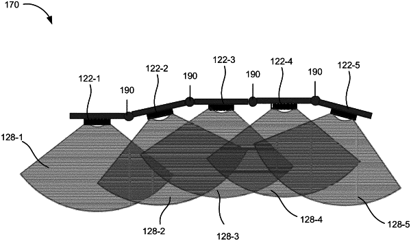
a plurality of transducers located in a housing configured to be taped or adhered to a portion of a body or located on a flexible strip configured to be taped or adhered to the portion of the body, and configured to:
transmit ultrasound signals directed to a target blood vessel, and
receive echo information associated with the transmitted ultrasound signals;
at least one processing device configured to:
process the echo information and generate a plurality of ultrasound images of the target blood vessel,
generate an estimated diameter of the target blood vessel at a plurality of locations based on the plurality of ultrasound images,
output image information associated with the target blood vessel based on the plurality of ultrasound images, and
output at least one of a maximum estimated diameter of the target blood vessel or the estimated diameters at the plurality of locations based on the image information; and
a plurality of position sensors or encoders located in the housing or on the flexible strip, where each of the plurality of transducers is located adjacent one of the plurality of position sensors or encoders, and
wherein when outputting image information associated with the target blood vessel, the at least one processing device is further configured to:
determine a relative position associated with each of the plurality of ultrasound images based on data from the plurality of position sensors or encoders, and
combine and rotate at least some of the plurality of ultrasound images to generate an image of the target blood vessel based on the determined relative position associated with each of the plurality of ultrasound images.
|