| CPC A61B 5/4509 (2013.01) [A61B 8/0875 (2013.01); A61B 8/14 (2013.01); A61B 8/5207 (2013.01); A61B 8/5223 (2013.01); G01S 15/8952 (2013.01); G06T 7/0012 (2013.01); A61B 8/485 (2013.01)] | 17 Claims |

|
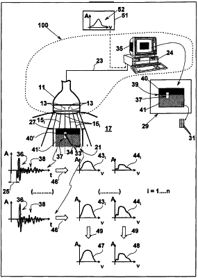
1. An apparatus (100) for assessing a condition of bone tissue in a patient's bone region (21), said apparatus (100) comprising:
an ultrasound device provided with an ultrasound probe (11) having an array of piezoelectric crystals that is configured to emit ultrasound pulses having a nominal frequency set between 2 and 9 MHz along a plurality of ultrasound propagation lines (15i) arranged in a sonographic plane (17) forming an ultrasound signal that can reach said patient's bone region (21), and to receive first raw ultrasound signals (36) reflected from a cortical part (40′) of said patient's bone region (21) and second raw ultrasound signals (38) reflected from a trabecular part (41′) of said patient's bone region (21), in response to said ultrasound pulses,
said ultrasound probe (11) is arranged to transmit said ultrasound pulses and to receive said first raw ultrasound signals (36) reflected from the cortical part (40′) of said patient's bone region (21) and said second raw ultrasound signals (38) reflected from the trabecular part (41′) from a same side of said patient's bone region (21);
a computer (24) configured to form a sonographic image (29) of a plane cross section of the bone region (21), taken along the sonographic plane (17) starting from said first raw ultrasound signals (36) reflected from the cortical part (40′) of said bone region (21) and said second raw ultrasound signals (38) reflected from the trabecular part (41′) of said bone region (21), said computer (24) is configured for displaying said sonographic image (29), that identifies a zone of said bone region to be investigated;
wherein said computer (24) is configured to receive and process said first raw ultrasound signals (36) reflected from the cortical part (40′) of said bone region (21), and said second raw ultrasound signals (38) reflected from the trabecular part (41′) of said bone region (21);
wherein said computer (24) is configured to separate said first raw ultrasound signals (36) reflected from the cortical part (40′) of said bone region (21) from said second raw ultrasound signals (38) reflected from the trabecular part (41′) of said bone region (21);
wherein said computer (24) is configured for extracting, starting from said first raw ultrasound signals (36) reflected from the cortical part (40′) of said bone region, a plurality of frequency spectra (43i, 47) associated with said cortical part (40′) and for extracting, starting from said second raw ultrasound signals (38) reflected from the trabecular part (41′) of said bone region (21), a plurality of frequency spectra (44i, 48) associated with said trabecular part (41′);
wherein said computer (24) is configured to memorize:
at least one healthy reference spectrum (52) associated with at least one healthy patient,
at least one intermediate reference spectrum associated with an intermediately-healthy patient,
at least one pathological reference spectrum associated with a pathological patient;
and wherein said computer (24) is configured to compare said plurality of frequency spectra (43i, 47) associated with said cortical part (40′) and/or said plurality of frequency spectra (44i, 48) associated with said trabecular part (41′) with said:
at least one healthy reference spectrum (52) associated with at least one healthy patient,
at least one intermediate reference spectrum associated with an intermediately-healthy patient,
at least one pathological reference spectrum associated with a pathological patient;
for determining the value of a diagnostic parameter according to said comparison.
|