| CPC G02B 21/088 (2013.01) [G02B 21/367 (2013.01); G06T 7/13 (2017.01); G06V 10/28 (2022.01); G06V 10/507 (2022.01); G06V 20/69 (2022.01); G06T 2207/10056 (2013.01); G06T 2207/30024 (2013.01)] | 10 Claims |

|
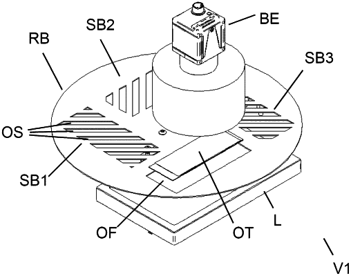
1. An apparatus for identifying respective cover slip regions (DGB) of respective cover slips having respective tissue sections (GS, GS1, GS2, GS3) on a specimen slide (OT), which has multiple optical identifiers (KE1, KE2, KE3), including
a planar light source (L),
an image acquisition unit (BE),
a holding unit (H) for positioning the specimen slide (OT) between the planar light source (L) and the image acquisition unit (BE),
a rotatable screen (RB) comprising at least one of a slit diaphragm (SB, SB1) and an opening (OF), wherein the slit diaphragm (SB, SB1) is comprised of multiple opening slits (OS),
the rotatable screen reversibly positionable between the planar light source (L) and the specimen slide (OT), such that the slit diaphragms (SB, SB1) and opening (OF) are reversibly positionable between the planar light source (L) and the specimen slide (OT),
an illumination unit (BL) designed to illuminate that surface (OFL) of the specimen slide (OT) which faces toward the image acquisition unit (BE),
and furthermore a control unit (K), which is designed,
in a first operating state, to activate the planar light source (L) and to acquire a completely illuminated transmitted light image (DB, DB2) of the specimen slide (OT) while upon the opening (OF) of the rotatable screen (RB) via the image acquisition unit (BE),
furthermore, in a second operating state, to activate the illumination unit (BL) and to acquire an incident light image (AB, AB2) of the specimen slide (OT) via the image acquisition unit (BE),
wherein the control unit is characterized in that,
furthermore, in a third operating state, to actuate the rotatable screen (RB) in such a way that the slit diaphragm (SB, SB1) is positioned between the planar light source (L) and the specimen slide (OT), and to activate the planar light source (L) and to acquire a partially darkened transmitted light image (PDB3) of the specimen slide (OT) via the image acquisition unit (BET),
to furthermore determine, on the basis of the incident light image (AB, AB2), respective spatial positions (P1, P2, P3) of respective optical identifiers (KE1, KE2, KE3),
furthermore, on the basis of the partially darkened transmitted light image (PDB3), to determine respective spatial locations of the respective cover slip regions (DGB),
furthermore, on the basis of the completely illuminated transmitted light image (DB, DB2), to determine respective spatial locations of the respective tissue sections (GS, GS1, GS2, GS3)
and, on the basis of the respective spatial positions (P1, P2, P3) of the respective optical identifiers (KE1, KE2, KE3), on the basis of the respective spatial locations of the respective cover slip regions (DGB), and on the basis of the respective spatial locations of the respective tissue sections (GS1, GS2, GS3), to assign the respective tissue sections to the respective optical identifiers (KE1, KE2, KE3).
|