| CPC A61B 3/102 (2013.01) [A61B 3/0041 (2013.01); A61B 90/39 (2016.02); G06T 7/60 (2013.01); A61B 2090/3904 (2016.02); G06T 2207/10101 (2013.01); G06T 2207/30041 (2013.01)] | 16 Claims |
|
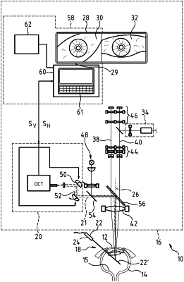
1. A system with an interface for providing visualization data for visualizing at least one section of a patient's eye,
comprising an OCT device for capturing OCT scanning data by scanning the section of the patient's eye by means of an OCT scanning beam and
comprising a computer unit for processing the OCT scanning data into the visualization data within the scope of an image rectification algorithm, which is designed to output the visualization data at the interface,
wherein the computer unit contains a view generation algorithm for calculating image data in relation to a view of a reference object arranged in the section of the patient's eye from geometry data about the reference object fed to the view generation algorithm and from the OCT scanning data obtained in relation to the reference object, and
wherein the computer unit has an algorithm control routine which specifies the image rectification algorithm and determines the image rectification algorithm from the image data of the view of the reference object calculated in the view generation algorithm and from OCT scanning data obtained in relation to the reference object by scanning the section of the patient's eye, said image rectification algorithm determining a rectification mapping which maps points of an image representation of the reference object in the OCT scanning data onto points of a generated view of the reference object which is based on image data calculated in relation to a view of a reference object arranged in the section of the patient's eye from geometry data about the reference object and which are fed to the view generation algorithm.
|