| CPC G06T 7/521 (2017.01) [A61B 1/00006 (2013.01); A61B 1/00194 (2022.02); A61B 1/05 (2013.01); A61B 1/0605 (2022.02); A61B 1/0638 (2013.01); A61B 90/36 (2016.02); A61B 1/000094 (2022.02)] | 20 Claims |
|
1. A surgical system, comprising:
a display;
a visualization system configured to scan the anatomy of a patient, comprising:
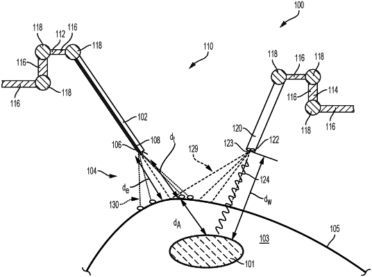
a structured light emitter configured to emit a structured pattern of electromagnetic radiation onto an anatomical structure;
a spectral light emitter configured to emit electromagnetic radiation comprising a plurality of wavelengths, wherein at least one wavelength of the plurality of wavelengths is selected to penetrate a portion of the anatomical structure and reflect off a subject tissue; and
an image sensor configured to detect the structured pattern of electromagnetic radiation reflected off the anatomical structure and the at least one wavelength reflected off the subject tissue; and
a surgical hub operably coupled to the visualization system and the display, wherein the surgical hub comprises a processor and a memory communicatively coupled to the processor, wherein the memory stores instructions executable by the processor to:
generate visualization data based on the scanned anatomy;
identify anatomical organs of the patient based on the generated visualization data;
generate virtual 3D constructs of the anatomical organs based on the generated visualization data;
display the virtual 3D constructs of the anatomical organs on the display;
identify anatomical structures based on the generated visualization data;
generate virtual 3D constructs of the anatomical structures based on the generated visualization data;
display the virtual 3D constructs of the anatomical structures on the display; and
overlay a surgical procedure plan on the display.
|