| CPC G06T 7/0012 (2013.01) [G01R 33/50 (2013.01); G01R 33/5608 (2013.01); G01R 33/56341 (2013.01); G06T 7/11 (2017.01); G06T 2207/10028 (2013.01); G06T 2207/10092 (2013.01); G06T 2207/20024 (2013.01); G06T 2207/20084 (2013.01); G06T 2207/20221 (2013.01); G06T 2207/30016 (2013.01); G06T 2207/30096 (2013.01)] | 13 Claims |
|
1. A system for mapping brain perivascular spaces, comprising:
a memory to store one or more images of a brain of a patient;
a processor coupled to the memory and configured to:
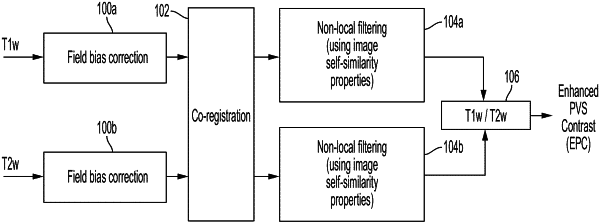
obtain a first image of the brain of the patient, the first image including a first contrast,
obtain a second image of the brain of the patient, the second image including a second contrast,
combine the first image and the second image to form a combined image, the combined image preserving and magnifying structures including the brain perivascular spaces within the combined image of the brain of the patient, the combined image including a third contrast, the third contrast being higher than the first contrast and the second contrast,
determine the brain perivascular spaces within the combined image of the brain of the patient, and
generate a three-dimensional (3-D) map of the brain perivascular spaces within the brain of the patient; and
a display configured to display the perivascular spaces of the brain of the patient to an operator.
|