| CPC G01N 21/6458 (2013.01) [G01N 1/30 (2013.01); G01N 21/76 (2013.01); G01N 33/54373 (2013.01)] | 85 Claims |

|
1. A method of enhancing a homogenous detection of an analyte in a cell that is in a sample, comprising:
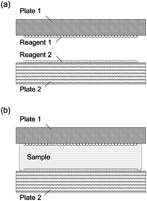
(a) providing a first plate and a second plate, each has a sample contact area on its surface, wherein the sample contact surfaces contact a sample comprising a cell that contains or is suspected of containing an analyte,
(b) providing a detection probe that specifically binds the analyte and is capable of emitting a light at a wavelength, wherein the detection probe diffuses in the sample;
(c) providing an optical enhancer that diffuses in the sample, binds to the cell, and is capable of emitting a light of a wavelength that overlaps with or within a 150 nm from the wavelength of the light emitted by the detection probe;
(d) sandwiching the sample, the detection probe, and the optical enhancer between the two sample contact areas of the two plates to form a thin layer of a thickness of 200 microns (um) or less; and
(e) imaging, using an imager, after step (d) and without using any washing step, the thin layer to detect the cell that has the analyte bound to the detection probe;
wherein the thin layer has a thickness that is selected so that for a given concentration of the cell in the thin layer, each individual cell does not substantially overlap other cells in the imaging;
wherein the thickness of the thin layer, the concentration of the detection probe in the sample, or the concentration of the optical enhancer in the sample is selected to make, in step (e) of imaging, in the thin layer, the location having a bound detection probe is distinguishable from the locations not having the bound detection probe, wherein the bound detection probe is the detection probe bound to the analyte in the cell.
|