| CPC A61B 1/000095 (2022.02) [A61B 1/0005 (2013.01); A61B 1/00078 (2013.01); A61B 1/000096 (2022.02); A61B 1/00103 (2013.01); A61B 1/00105 (2013.01); A61B 1/043 (2013.01); A61B 1/05 (2013.01); A61B 1/0684 (2013.01); A61B 5/0035 (2013.01); A61B 5/0071 (2013.01); A61B 5/0086 (2013.01); A61B 5/7267 (2013.01); A61B 5/7425 (2013.01); A61M 31/005 (2013.01); G06T 5/00 (2013.01); G06T 7/30 (2017.01); H04N 5/265 (2013.01); H04N 5/2624 (2013.01); H04N 5/272 (2013.01); H04N 23/45 (2023.01); H04N 23/54 (2023.01); H04N 23/74 (2023.01); G06T 2207/10048 (2013.01); G06T 2207/10068 (2013.01); G06T 2207/10152 (2013.01); G06T 2207/20081 (2013.01); G06T 2207/20221 (2013.01); H04N 23/555 (2023.01)] | 20 Claims |
|
1. An endoscope, comprising:
one or more image sensors designed to capture image data in visible light and image data in a nonvisible portion of the electromagnetic spectrum, sensels of the one or more image sensors having overlapping wavelength sensitivity bands;
an insertion shaft designed to support the one or more image sensors at or near a distal tip, the insertion shaft and support designed to support the one or more image sensors for endoscopic insertion to a surgical site in a human body;
a plurality of light emitting diodes (LEDs) of different wavelengths, designed to deliver illumination to the surgical site, including at least one LED to emit in visible wavelengths and at least one wavelength to emit at one or more wavelengths designed to induce fluorescence;
illumination intensity of the LEDs and sensor sensitivity of the image sensors being designed to compensate for each other; and
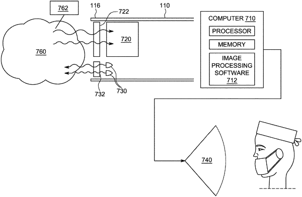
image processing software trained through machine learning to enhance image quality of at least the nonvisible portion of the image, including by subtraction of values of one sensel from another with an overlapping wavelength sensitivity band to recover an image value in the wavelength range of the non-overlap, and to present the enhanced nonvisible image as a real-time, visible presentation to a surgeon.
|