| CPC G06T 7/0016 (2013.01) [A61B 5/02007 (2013.01); A61B 5/7264 (2013.01); A61B 5/7275 (2013.01); A61B 5/743 (2013.01); G06T 7/38 (2017.01); G06T 11/008 (2013.01); G16H 30/40 (2018.01); G16H 50/30 (2018.01); G16H 50/50 (2018.01); G06T 2207/20081 (2013.01); G06T 2207/30104 (2013.01)] | 19 Claims |

|
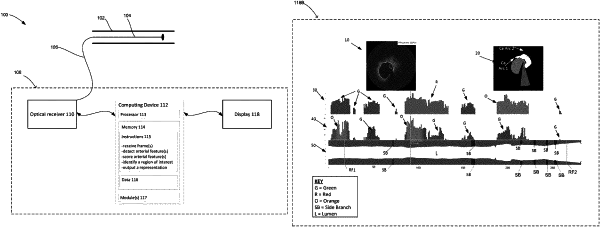
1. A method of displaying one or more arterial features relative to at least one of a first pullback representation or a second pullback representation, the method comprising:
receiving, by one or more processors, a first group of intravascular frames obtained from a first pullback pre-percutaneous coronary intervention;
detecting, by the one or more processors based on a determined calcium arc or a determined calcium volume, a calcium burden in each frame of the first group of intravascular frames;
scoring, by the one or more processors, the calcium burden in each frame of the first group of intravascular frames;
predicting, by the one or more processors executing a machine learning model, based on the scored calcium burden, stent expansion for a region of interest, wherein:
at least one input into the machine learning model is the scored calcium burden; and
outputting, by the one or more processors, a two-dimensional representation of the first group of intravascular frames, wherein:
the two-dimensional representation is symmetrical about a longest axis of the two-dimensional representation,
the output includes a visual indication corresponding to the predicted stent expansion for the region of interest, and
the visual indication is a severity indicator for stent over or under expansion.
|