| CPC G06T 7/0012 (2013.01) [A61B 8/0891 (2013.01); A61B 8/12 (2013.01); G06T 7/11 (2017.01); G06T 7/70 (2017.01); G16H 50/30 (2018.01); G06T 2207/10132 (2013.01); G06T 2207/20081 (2013.01); G06T 2207/30101 (2013.01)] | 19 Claims |
|
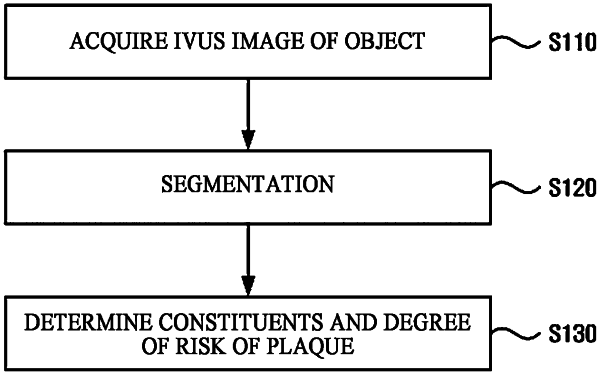
1. A method of performing analysis of a cardiovascular image of a subject, comprising:
obtaining first image for at least part of a cardiovascular system of the subject; and
obtaining second image by processing the first image based on a diagnosis assistance model, the diagnosis assistance model being trained based on first blood vessel image and second blood vessel image corresponding to the first blood vessel image;
wherein the first image and the first blood vessel image are taken with a same imaging method,
wherein the second image is generated based on the first image to provide analysis information on at least a partial region of the first image,
wherein a location of a cardiovascular vessel in the second blood vessel image corresponds to a location of a cardiovascular vessel in the first blood vessel image,
wherein the first blood vessel image is divided into a plurality of first rectangular regions rotated at a predetermined angle, and
wherein the diagnosis assistance model is trained based on the first rectangular regions of the first blood vessel image.
|