| CPC G16H 50/20 (2018.01) [G06T 7/0012 (2013.01); G06T 7/10 (2017.01); G06V 20/695 (2022.01); G16H 10/40 (2018.01); G16H 30/20 (2018.01); G16H 30/40 (2018.01); G16H 50/30 (2018.01); G06T 2207/10024 (2013.01); G06T 2207/10048 (2013.01); G06T 2207/30004 (2013.01); G06T 2207/30024 (2013.01)] | 21 Claims |

|
1. A method comprising:
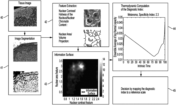
receiving, by a processor, data representing at least one cell that has been identified in an area of an image and segmented into a nuclear area and a cellular area;
calculating, by the processor for the at least one cell and based at least in part on the data, a plurality of information surface values each derived as a function of both:
(i) a nuclear contrast feature, wherein the nuclear contrast feature comprises a temperature difference between the nuclear area and the cellular area; and
(ii) a nuclear area feature, wherein the nuclear area feature comprises a ratio of the nuclear area to a nuclear volume projection;
calculating, by the processor, a diagnostic score based at least in part on the plurality of information surface values; and
determining, by the processor based at least in part on the diagnostic score, a normality status of the area of the image.
|