| CPC G02B 21/367 (2013.01) [G02B 21/0064 (2013.01); G06N 3/045 (2023.01); G06T 7/0012 (2013.01); G06T 2207/10056 (2013.01); G06T 2207/20081 (2013.01); G06T 2207/30096 (2013.01)] | 10 Claims |

|
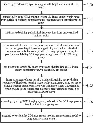
1. A margin assessment method, comprising:
selecting a predetermined specimen region with a target lesion from a skin of a subject;
extracting, by using a harmonic generation microscopy (HGM) imaging system, a plurality of 3D image groups within a range from a surface of a plurality of positions in the predetermined specimen region to a predetermined depth, wherein each of the 3D image groups includes a series of a plurality of 2D images;
obtaining and staining a plurality of pathological tissue sections from the predetermined specimen region;
examining the plurality of pathological tissue sections to generate a plurality of pathological results and define a margin of the target lesion, using the plurality of pathological results as a plurality of standard examination results that correspond to the 3D image groups according to the plurality of positions, and labeling the 3D image groups to generate a plurality of labeled 3D image groups;
pre-processing the plurality of labeled 3D image groups and dividing the plurality of labeled 3D image groups into a training set, a validation set, and a test set;
fitting parameters of a deep learning model with the training set, predicting responses of the fitted deep learning model with the validation set, and then using the test set to evaluate whether a final model fitted with the training set meets a predetermined condition, wherein the final model that meets the predetermined condition is taken as a margin assessment model;
extracting, by using the HGM imaging system, a plurality of to-be-identified 3D image groups from a plurality of locations in a target region; and
inputting the to-be-identified 3D image groups into the margin assessment model to generate assessment results.
|