| CPC A61C 9/006 (2013.01) [A61B 1/00096 (2013.01); A61B 1/00172 (2013.01); A61B 1/00193 (2013.01); A61B 1/24 (2013.01); G02B 27/4222 (2013.01); G02B 27/4227 (2013.01); G06T 7/521 (2017.01); G06T 7/586 (2017.01); G06T 7/80 (2017.01); G06T 7/85 (2017.01); G06T 17/00 (2013.01); H04N 9/3161 (2013.01); G06T 2207/10024 (2013.01); G06T 2207/10028 (2013.01); G06T 2207/10052 (2013.01); G06T 2207/30036 (2013.01)] | 29 Claims |

|
1. An apparatus for intraoral scanning, the apparatus comprising:
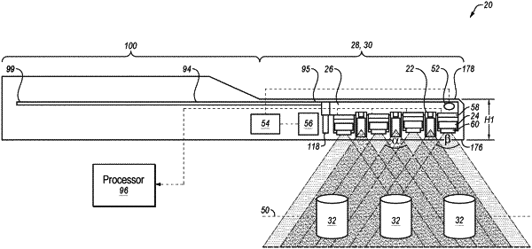
an elongate handheld wand comprising a probe at a distal end of the elongate handheld wand;
a plurality of structured light projectors disposed in the probe at the distal end of the elongate handheld wand, each structured light projector comprising at least one light source and a pattern generating optical element, wherein each structured light projector is configured to project a pattern of light defined by a plurality of projector rays onto an intraoral surface by transmitting light from the at least one light source through the pattern generating optical element that uses at least one of diffraction or refraction to generate the pattern of light when the light source is activated, wherein each of the plurality of structured light projectors is to project respective distributions of discrete unconnected spots of light on the intraoral surface simultaneously or at different times, wherein each of the plurality of projector rays corresponds to a spot of the discrete unconnected spots of light, wherein the pattern of light comprises a non-coded structured light pattern, and wherein the discrete unconnected spots of light comprise an approximately uniform distribution of discrete unconnected spots of light;
two or more cameras disposed in the probe at the distal end of the elongate handheld wand, each of the two or more cameras comprising a camera sensor having an array of pixels, wherein each of the two or more cameras is configured to capture a plurality of images that depict at least a portion of the projected pattern of light on an intraoral surface;
at least one uniform light projector configured to project white light onto the intraoral surface, wherein at least one of the two or more cameras is configured to capture two-dimensional color images of the intraoral surface using illumination from the uniform light projector; and
one or more processors configured to:
drive the plurality of structured light projectors to project the pattern of light and the two or more cameras to capture the plurality of images;
receive the plurality of images that depict at least the portion of the projected pattern of light on the intraoral surface from the two or more cameras;
determine a correspondence between points in the pattern of light and points in the plurality of images that depict at least the portion of the projected pattern of light on the intraoral surface by:
accessing calibration data that associates camera rays corresponding to pixels on the camera sensor of each of the two or more cameras to projector rays of the plurality of projector rays that are generated when the light is transmitted from the light source through the pattern generating optical element; and
determining intersections of projector rays and camera rays corresponding to at least the portion of the projected pattern of light using the calibration data, wherein intersections of the projector rays and the camera rays are associated with three-dimensional points in space;
identify three-dimensional locations of the projected pattern of light based on agreements of the two or more cameras on there being the projected pattern of light by projector rays at certain intersections; and
use the identified three-dimensional locations to generate a digital three-dimensional model of the intraoral surface.
|