| CPC A61B 6/504 (2013.01) [A61B 5/0066 (2013.01); A61B 5/0075 (2013.01); A61B 5/055 (2013.01); A61B 5/7267 (2013.01); A61B 5/742 (2013.01); A61B 5/7475 (2013.01); A61B 6/032 (2013.01); A61B 6/037 (2013.01); A61B 6/463 (2013.01); A61B 6/467 (2013.01); A61B 6/481 (2013.01); A61B 6/5205 (2013.01); A61B 8/12 (2013.01); A61B 8/14 (2013.01); A61K 49/04 (2013.01); G06F 18/10 (2023.01); G06T 7/0012 (2013.01); G06V 10/20 (2022.01); G06V 10/245 (2022.01); G06V 10/761 (2022.01); G06V 10/764 (2022.01); G06V 10/82 (2022.01); G06T 2207/10081 (2013.01); G06T 2207/10088 (2013.01); G06T 2207/10101 (2013.01); G06T 2207/10132 (2013.01); G06T 2207/20081 (2013.01); G06T 2207/30048 (2013.01); G06T 2207/30101 (2013.01); G06V 10/247 (2022.01)] | 26 Claims |

|
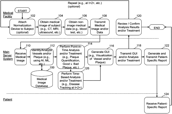
1. A system for tracking a plaque-based disease based on non-invasive medical image analysis of a medical image of a subject to facilitate determination of a state of plaque progression, regression, or stabilization, the system comprising:
one or more computer readable storage devices configured to store a plurality of computer executable instructions; and
one or more hardware computer processors in communication with the one or more computer readable storage devices and configured to execute the plurality of computer executable instructions in order to cause the system to:
access a first set of plaque parameters of one or more vessels of a subject, wherein the first set of plaque parameters are derived from a first medical image of the subject comprising the one or more vessels, the first medical image of the subject obtained non-invasively at a first point in time, wherein the first set of plaque parameters comprises total plaque volume, non-calcified plaque volume, and calcified plaque volume identified in the one or more vessels of the subject at the first point in time;
access a second medical image of the subject, the second medical image of the subject obtained non-invasively at a second point in time, the second point in time being later than the first point in time, wherein the second medical image of the subject comprises the one or more vessels of the subject;
analyze the second medical image to generate a second set of plaque parameters of the one or more vessels of the subject, wherein the second set of plaque parameters comprises total plaque volume, non-calcified plaque volume, and calcified plaque volume identified in the one or more vessels of the subject at the second point in time;
determine a change between one or more of the first set of plaque parameters and one or more the second set of plaque parameters; and
generate a display of the determined change between one or more of the first set of plaque parameters and one or more of the second set of plaque parameters to facilitate determination of the state of plaque progression, regression, or stabilization based at least part on the determined change.
|