| CPC G02B 23/2469 (2013.01) [A61B 1/00043 (2013.01); A61B 1/000094 (2022.02); A61B 1/044 (2022.02); A61B 1/05 (2013.01); A61B 1/0646 (2013.01); A61B 1/0655 (2022.02); A61B 1/07 (2013.01)] | 7 Claims |

|
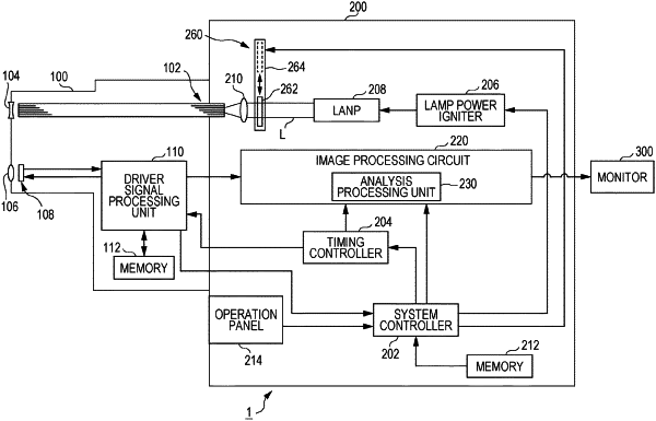
1. An endoscope system capable of operating in a normal observation mode for irradiating a biological tissue with white light to acquire a first image and a special observation mode for irradiating the biological tissue with light of a specific wavelength band to acquire a second image, comprising:
a lamp that irradiates the biological tissue with illumination light including at least red light R1 of a first wavelength band, green light G1 of a second wavelength band, blue light B1 of a third wavelength band, red light R2 of a fourth wavelength band, green light G2 of a fifth wavelength band, and blue light B2 of a sixth wavelength band;
an image sensor that generates image data based on reflected light from the biological tissue generated by irradiating the biological tissue with the illumination light, the image data including a first RGB image, a second RGB image different from the first RGB image, and a correction image;
an image processor that acquires the image data including the first RGB image, the second RGB image, and the correction image from the image sensor and performs a predetermined image process; and
a display that displays a special light image generated by the predetermined image process of the image processor on a screen,
wherein:
at least the second wavelength band, the third wavelength band, the fifth wavelength band, and the sixth wavelength band are defined with boundaries therebetween,
the boundaries are wavelengths at isosbestic points at which transmittance becomes constant regardless of oxygen saturation,
the second wavelength band includes isosbestic points other than the isosbestic points,
the first RGB image of the image data includes R1 image data corresponding to R1 of the first wavelength band, G1 image data corresponding to G1 of the second wavelength band, and B1 image data corresponding to B1 of the third wavelength band,
the second RGB image of the image data includes R2 image data corresponding to R2 of the fourth wavelength band, G2 image data corresponding to G2 of the fifth wavelength band, and B2 image data corresponding to B2 of the sixth wavelength band,
the correction image is used as a reference when the image processor corrects RGB values of the first RGB image and the second RGB image,
the image processor generates the special light image by performing an image process using the G1 image data and at least one of the R1 image data, the B1 image data, the R2 image data, the G2 image data, and the B2 image data,
the first wavelength band is 630±3 nm to 700±3 nm,
the second wavelength band is 524±3 nm to 582±3 nm,
the third wavelength band is 452±3 nm to 502±3 nm,
the fourth wavelength band is 582±3 nm to 630±3 nm,
the fifth wavelength band is 502±3 nm to 524±3 nm,
the sixth wavelength band is from 420±3 nm to 452±3 nm,
452±3 nm, 502±3 nm, 524±3 nm, and 582±3 nm are wavelengths at the isosbestic points,
the image processor assigns the G2 image data to a blue wavelength region, and assigns the R2 image data to a green wavelength region to generate a blood transparentized image in which the blood is transparent,
the image processor multiplies the G1 image data by a predetermined subtraction parameter coefficient, and
the display displays the blood transparentized image on the screen.
|