| CPC A61B 8/463 (2013.01) [A61B 8/0858 (2013.01); A61B 8/14 (2013.01); A61B 8/483 (2013.01); A61B 8/485 (2013.01); A61B 8/5207 (2013.01); A61B 8/5253 (2013.01); A61B 8/5276 (2013.01)] | 19 Claims |

|
1. An ultrasound imaging system comprising:
an ultrasound imaging device configured to generate shear wave signals responsive to shear wave tracking echoes received by an ultrasound probe communicatively coupled to the ultrasound imaging device; and
a processor integral with or communicatively coupled to the ultrasound imaging device, wherein the processor includes:
a shear wave processor configured to calculate tissue stiffness values based, at least in part, on the shear wave signals;
a confidence map generator configured to calculate confidence values based on at least one confidence factor; and
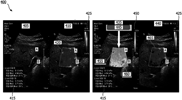
an image processor configured to generate an ultrasound image including a graphical overlay of tissue stiffness values for one or more pixels within a region of interest and a confidence map, wherein the confidence map is configured to provide, based on the calculated confidence values, an indication of reliability of the tissue stiffness values within the region of interest,
wherein the confidence map represents different confidence values with different colors.
|