| CPC A61B 8/06 (2013.01) [A61B 5/7267 (2013.01); A61B 8/0891 (2013.01); A61B 8/463 (2013.01); A61B 8/469 (2013.01); A61B 8/488 (2013.01); A61B 8/5223 (2013.01); G01F 1/667 (2013.01); G16H 15/00 (2018.01); G16H 30/20 (2018.01); G16H 40/60 (2018.01); G16H 50/20 (2018.01); G16H 50/50 (2018.01); A61B 8/0866 (2013.01); A61B 8/5246 (2013.01)] | 6 Claims |

|
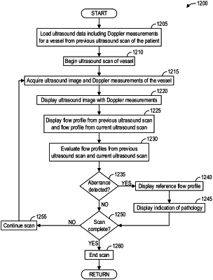
1. A method, comprising:
acquiring, via an ultrasound probe, an ultrasound image of a vessel;
acquiring, via the ultrasound probe, Doppler measurements of blood flow within the vessel;
displaying, via a display device, a graphical user interface including the ultrasound image with data of the Doppler measurements, and a flow profile constructed from the Doppler measurements of blood flow within the vessel;
detecting, with a detection module, an abnormality in the flow profile, wherein the flow profile is input to the detection module as an image or as a time series of the flow profile; and
adjusting the display of the graphical user interface to indicate the abnormality via displaying, via the display device, a reference flow profile for the vessel superimposed over the flow profile in the graphical user interface, wherein adjusting the display includes one or more of time-aligning a peak of the reference flow profile with a peak of the flow profile, and scaling the reference flow profile to match an amplitude of the peak of reference flow profile with an amplitude of the peak of the flow profile.
|