| CPC A61B 6/507 (2013.01) [A61B 6/027 (2013.01); A61B 6/032 (2013.01); A61B 6/504 (2013.01); A61B 6/5205 (2013.01); G06T 7/38 (2017.01); G06T 11/003 (2013.01); G06T 11/008 (2013.01); G06T 2207/10081 (2013.01); G06T 2207/20081 (2013.01); G06T 2207/30016 (2013.01); G06T 2207/30104 (2013.01)] | 20 Claims |
|
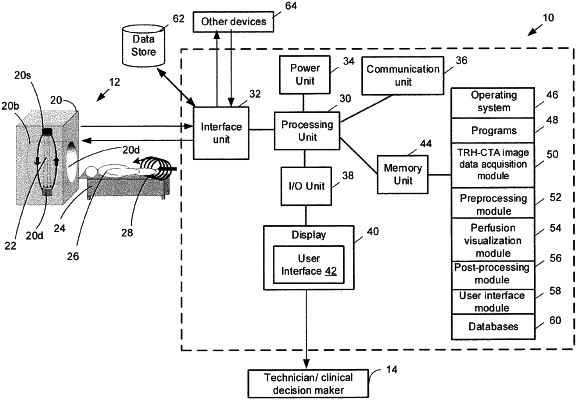
1. A system for providing at least one Computed Tomography Angiography (CTA) perfusion functional map, wherein the system comprises:
at least one processor that is configured to:
obtain Time Resolved Helical CTA (TRH-CTA) image data;
preprocess the TRH-CTA helical image data to generate preprocessed TRH-CTA helical image data;
generate time density curve data for a plurality of voxels from the preprocessed TRH-CTA helical image data for an axial imaging slice, where the time density curve data comprise intensity values for different phases of the preprocessed TRH-CTA helical image data arranged sequentially in time;
generate the at least one perfusion functional map for the axial imaging slice by at least one of: (1) applying at least one mapping function to different phases of the time density curve data corresponding to the axial imaging slice; (2) applying a deconvolution method to the time density curve data; and (3) applying a non-deconvolution method to the time density curve data; and
perform filtering in the spatial domain or the frequency domain on the at least one perfusion functional map; and
a display that is coupled to the at least one processor for receiving and displaying the at least one filtered perfusion functional map.
|