| CPC G16H 30/40 (2018.01) | 15 Claims |

|
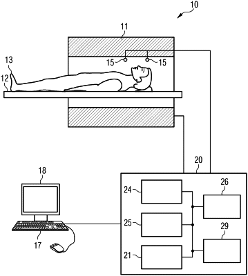
1. A magnetic resonance (MR) imaging system for generating an image of an examination object, the MR imaging system comprising:
magnetic field gradient circuitry having a component property which, to generate the image of the examination object, sets a value in a first value range established by the MR imaging system,
wherein a further magnetic field gradient circuitry of a further MR system has a corresponding component property with a second value range established by the further MR imaging system; and
gradient control circuitry configured to control the magnetic field gradient circuitry to generate the image of the examination object by operating in a compatibility mode in which the gradient control circuitry executes a scan protocol to only allow values to be set for the component property of the magnetic field gradient circuitry that lie within an overlap range that represents values of the first value range and the second value range that overlap with one another,
wherein the component property of the magnetic field gradient circuitry includes values associated with a gradient field strength and a gradient rate of rise of the magnetic field gradients generated by the MR imaging system via the magnetic field gradient circuitry,
wherein the gradient control circuitry and the further magnetic field gradient circuitry of the MR system and the further MR system, respectively, execute the same scan protocol when operating in respective compatibility modes such that the image of the examination object generated via the MR imaging system has the same image appearance as a further image of the examination object generated via the further MR imaging system, and
wherein the scan protocol that is executed in accordance with the compatibility mode via the MR system and the further MR system defines a gradient amplitude, a plateau time, a ramp time, and a fall time for an excitation pulse and a corresponding re-phasing pulse.
|