| CPC A61B 6/4411 (2013.01) [A61B 6/035 (2013.01); A61B 6/4452 (2013.01); A61B 6/5229 (2013.01)] | 18 Claims |
|
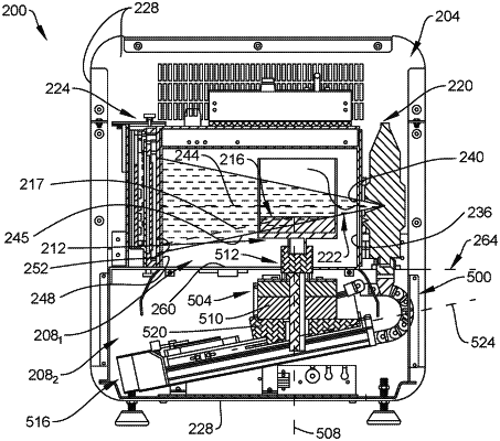
1. A method of imaging a tissue specimen in a cabinet including an x-ray source and an x-ray detector positioned along a first axis, the method comprising:
placing the tissue specimen on an object holder disposed within a chamber of the cabinet, the object holder being disposed at least partially between the x-ray source and the x-ray detector, wherein the object holder is selectively moveable in a direction along the first axis such that the object holder defines a first position;
determining a position of the tissue specimen at the first position of the object holder relative to the x-ray source and the first axis;
after the position of the tissue specimen at the first position is determined, moving the object holder within the chamber towards a second position based on the determined position of the tissue specimen;
rotating the object holder around a second axis perpendicular to the first axis, wherein the object holder rotates while in the second position;
simultaneously with rotating the object holder, emitting one or more x-ray beams along the first axis and through the tissue specimen;
generating a plurality of two-dimensional x-ray images of the tissue specimen; and
reconstructing the plurality of two-dimensional x-ray images into a three-dimensional data set of the tissue specimen.
|