| CPC A61B 8/0825 (2013.01) [A61B 8/0841 (2013.01); A61B 8/085 (2013.01); A61B 8/14 (2013.01); A61B 8/463 (2013.01); A61B 10/0233 (2013.01)] | 9 Claims |

|
1. A computing device, comprising:
at least one processor configured to be communicatively coupled to an ultrasound probe operated by a user; and
at least one memory storing computer-executable instructions that implement an ultrasound workflow tool for facilitating a diagnostic procedure, the computer-executable instructions when executed by the at least one processor cause the computing device to:
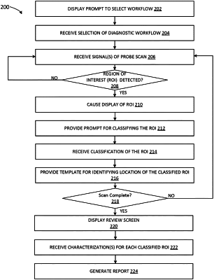
provide a first prompt for selection of a workflow from among a plurality of workflows;
receive, from the user, a selection of a selected workflow from among the plurality of workflows;
receive one or more signals from the ultrasound probe operated by the user while scanning an anatomical feature of a patient;
detect, by the ultrasound workflow tool, a region of interest associated with the scanned anatomical feature;
during detection of the region of interest, cause display of the region of interest on a display of the computing device;
during display of the region of interest, provide a second prompt for classifying the detected region of interest;
receive from the user a classification of the region of interest in response to the second prompt provided by the computing device;
in response to receiving the classification, provide a template for identifying at least one location of the classified region of interest, wherein:
providing the template comprises providing a schematic representation of the anatomical feature,
the schematic representation comprising a first pictogram comprising a first two-dimensional line diagram of the anatomical feature, the first pictogram comprising a plurality of numerically identified circular regions that radiate from a nipple of the anatomical feature, wherein the anatomical feature is a breast, and
the schematic representation comprising a second pictogram, the second pictogram comprising a second two-dimensional line diagram of the anatomical feature, the second pictogram comprising a plurality of alphabetically identified regions that radiate from a top surface of the breast;
receive a first signal, in response to a first user selection on the first pictogram, of a location in relation to the nipple of the classified region of interest;
receive a second signal, in response to a second user selection on the second pictogram, of a depth of the classified region of interest;
translate the first user selection and the second user selection into number-letter coordinates identifying the at least one location of the classified region of interest;
provide a review screen on the display comprising one or more fields for characterizing the classified region of interest;
receive from the user a description into each of the one or more fields for characterizing the classified region of interest; and
generate a report of the diagnostic procedure based in part on the classified region of interest, the identified at least one location, and the received one or more descriptions.
|