| CPC A61B 1/00193 (2013.01) [A61B 1/00009 (2013.01); A61B 1/00096 (2013.01); A61B 1/00174 (2013.01); A61B 1/00186 (2013.01); A61B 1/00194 (2022.02); G02B 23/2415 (2013.01)] | 19 Claims |

|
1. An endoscope apparatus comprising:
a first optical lens;
a second optical lens;
at least one image pickup device configured to generate:
a first image pickup signal based on a first optical image formed on the at least one image pickup device from the first optical lens, and
a second image pickup signal based on a second optical image formed on the at least one image pickup device from the second optical lens, wherein a first effective optical path length between the first optical lens and the at least one image pickup device is different than a second effective optical path length between the second optical lens and the at least one image pickup device; and
a processor comprising hardware, the processor being configured to:
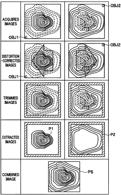
generate a first image of an object based on the first image pickup signal, the first image having a first focused area of the object due to the first effective optical path length;
generate a second image of an object based on the second image pickup signal, the second image having a second focused area of the object due to the second effective optical path length;
correct a parallax in at least one of the first image or the second image;
determine an area in the first image having a contrast higher than a contrast of an area in the second image to be included in the first focused area to extract only the first focused area from the first image;
determine an area in the second image having a contrast higher than a contrast of an area in the first image to be included in the second focused area to extract only the second focused area from the second image; and
combine the first focused area and the second focused area to generate an endoscope image different from each of the first image and the second image.
|