| CPC A61B 6/037 (2013.01) [A61B 6/0407 (2013.01); A61B 6/4258 (2013.01); A61B 6/4266 (2013.01); A61B 6/4275 (2013.01); A61B 6/4291 (2013.01); A61B 6/447 (2013.01); A61B 6/4429 (2013.01); A61B 6/544 (2013.01); A61B 6/547 (2013.01); A61B 6/589 (2013.01); G01T 1/1603 (2013.01); G21K 1/025 (2013.01); A61B 6/102 (2013.01); A61B 6/105 (2013.01); A61B 6/4417 (2013.01); A61B 6/4494 (2013.01); A61B 6/5205 (2013.01)] | 32 Claims |
|
1. A method of nuclear medicine imaging comprising:
providing a nuclear medicine tomography system comprising:
a support for a subject of a tomography procedure, the subject having a region of interest (ROI), said support having an axis;
a detector carrier, wherein at least one of said subject support and said detector carrier is movable axially relative to the other;
wherein said detector carrier is rotatable about said support;
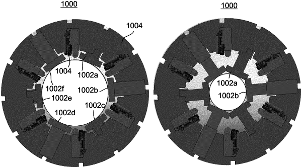
a plurality of detector heads mounted on said detector carrier, at least some of said detector heads each configured to be extended along a respective extension axis between said detector carrier and the ROI, from a first, most retracted position relative to said detector carrier, to a second position closer to the ROI than said first position, and to be retracted from said second position to said first position;
selecting a bore geometry for said tomography system, said bore geometry based on a part of the subject's body having the ROI;
linearly extending each of a plurality of said plurality of detector heads along a respective said extension axis from a respective said first position to respective said second position, based on said selected bore geometry;
rotating said detector carrier and acquiring images of said region of interest.
|