| CPC A61M 39/0208 (2013.01) [A61M 25/00 (2013.01); A61M 39/0247 (2013.01); A61B 2034/105 (2016.02); A61B 2034/107 (2016.02); A61B 2090/365 (2016.02); A61M 2039/0223 (2013.01); A61M 2039/0232 (2013.01); A61M 2039/0238 (2013.01); A61M 2205/32 (2013.01); A61M 2210/0687 (2013.01)] | 15 Claims |

|
1. A for accessing an intracranial subarachnoid space (ISAS) through a blood vessel wall of a patient to administer a therapeutic agent, the method comprising:
acquiring a 3D volumetric reconstruction of the vessel wall;
identifying a target location in the 3D reconstruction for accessing the ISAS through the vessel wall with a delivery catheter;
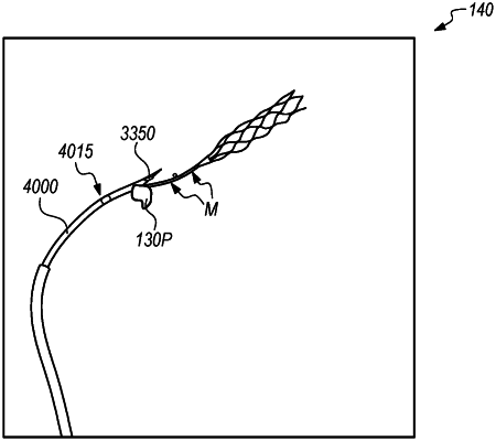
overlaying a portion of the 3D reconstruction including the target location on a fluoroscopy imaging display of the patient's anatomy including the vessel wall;
using the overlaid portion of the 3D reconstruction and fluoroscopy imaging display to visually track movement of the delivery catheter within the vessel to the target location;
penetrating the vessel wall at the target location to create an anastomosis between the vessel and the ISAS;
accessing the ISAS through the anastomosis with the delivery catheter; and
administering a therapeutic agent from the delivery catheter into the ISAS.
|