| CPC A61B 5/489 (2013.01) [A61B 5/0066 (2013.01); A61B 5/0075 (2013.01); A61B 5/02007 (2013.01); A61B 5/0068 (2013.01); A61B 5/0084 (2013.01); A61B 5/14546 (2013.01); A61B 5/418 (2013.01)] | 27 Claims |

|
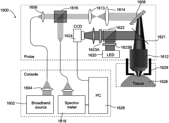
1. An imaging probe for locating a target blood vessel located under a portion of a tissue surface, the imaging probe located outside the portion of the tissue surface, the imaging probe comprising:
an objective lens;
a background imaging channel comprising a background light source configured to illuminate with background light a tissue region located under the portion of the tissue surface through the objective lens, using wide-field illumination comprising a background light component having a substantially high susceptibility to absorption by particles in said portion of the target blood vessel; and
an illuminating optical channel comprising an illuminating light source and an illumination light processing unit, respectively configured to illuminate with illuminating light and image through the objective lens, along a focal line within the tissue region;
wherein the illuminating optical channel also comprises an optical arrangement configured to direct light backscattered from the focal line to the illumination light processing unit;
wherein the illumination light processing unit is configured to decode position from illuminating light backscatter returning from the focal line, the focal line located under the portion of the tissue surface;
wherein the background light and the illuminating light both illuminate from the objective lens through an optically transparent surface of the imaging probe, wherein said imaging is through said optically transparent surface, and wherein said optically transparent surface is configured to be in contact with the portion of the tissue surface under which the target blood vessel is located; and
wherein, in addition to the illuminating optical channel which is dispersed across the focal line, said objective lens is adjustable relative to the optically transparent surface, to move an optical line of sight of confocal imaging of the background imaging channel and the illuminating optical channel, such that said imaging probe is configured to image at least a portion of the blood vessel located under the portion of the tissue surface.
|