| CPC A61B 1/043 (2013.01) [A61B 1/00009 (2013.01); A61B 1/00052 (2013.01); A61B 1/00096 (2013.01); A61B 1/00186 (2013.01); A61B 1/015 (2013.01); A61B 1/045 (2013.01); A61B 1/05 (2013.01); A61B 1/051 (2013.01); A61B 1/0607 (2013.01); A61B 1/0638 (2013.01); A61B 1/0655 (2022.02); A61B 1/126 (2013.01); A61B 1/307 (2013.01); A61B 5/0071 (2013.01); A61M 13/003 (2013.01); G02B 21/16 (2013.01); A61B 2562/0233 (2013.01); A61M 2210/1085 (2013.01)] | 10 Claims |

|
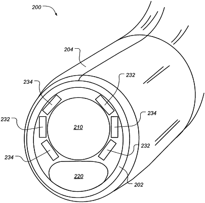
1. A single-use, disposable cannula for multi-band imaging of a patient's internal organ, said cannula comprising:
an imaging structure at a distal portion of the cannula;
a gas inlet port that is at a proximal portion of the cannula and is configured to receive insufflating gas, a gas outlet port at the distal portion of the cannula, and a gas conduit between said gas inlet port and gas outlet port, wherein said gas outlet port is configured to direct insufflating gas delivered thereto through said gas conduit in a flow over said imaging structure configured to clear the imaging structure;
a source of insufflating gas under pressure selectively coupled to said gas inlet port to supply insufflating gas thereto;
wherein said imaging structure at the distal portion of the cannula further comprises:
a light source configured to illuminate said internal organ with white light from a white LED and with non-white light from a narrow-band, non-white LED, wherein the white LED and the non-white LED illuminate the internal organ from different angles;
a multi-pixel, backside-illuminated, two-dimensional white light sensor array configured to image white light from the organ and to generate white light image data;
a multi-pixel, backside-illuminated, two-dimensional non-white sensor array configured to image non-white light from the organ and to generate non-white light image data; and
a readout circuit electrically and physically integrated with said white light sensor array and said non-white light sensor array into a circuit stack; and
wherein the white light image data and the non-white light image data represent respective white and non-white images of the organ that are both spatially and temporally registered with each other.
|