| CPC G06T 7/0012 (2013.01) [A61B 6/481 (2013.01); A61B 6/504 (2013.01); A61B 6/507 (2013.01); A61B 6/5217 (2013.01); G06N 3/08 (2013.01); G06N 20/00 (2019.01); G06T 15/08 (2013.01); G06T 2200/04 (2013.01); G06T 2200/08 (2013.01); G06T 2207/20081 (2013.01); G06T 2207/20084 (2013.01); G06T 2207/30048 (2013.01); G06T 2207/30101 (2013.01)] | 22 Claims |
|
12. A system for assessing a vessel obstruction, comprising:
memory configured to store program instructions and to store a volumetric image dataset for a target organ that includes a vessel of interest (VOI); and
at least one processor that, when executing the program instructions stored in the memory, is configured to:
extract an axial trajectory extending along the vessel of interest (VOI) within the volumetric image dataset;
create a three-dimensional (3D) multi-planer reformatted (MPR) image based on the volumetric image dataset and the axial trajectory of the VOI;
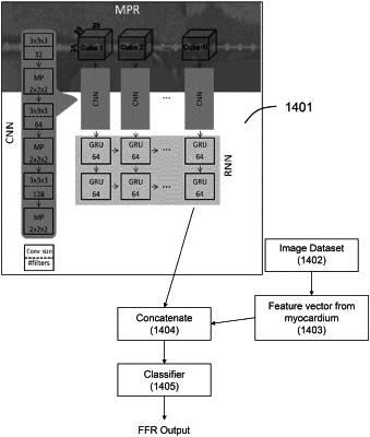
extract a VOI parameter along the vessel of interest from the MPR image utilizing a machine learning-based vessel obstruction assessment (VOA) model, wherein the VOI parameter represents functionally significant coronary lesion severity; and
output for display a visual representation of the VOI parameter along the VOI using a simulated angiographic image, wherein the simulated angiographic image is created from the volumetric image dataset under a given perspective;
wherein the machine learning-based VOA model automatically extracts image features associated with cubes generated from the MPR image utilizing a first machine learning-based model trained from image data with known labels, and automatically determines the VOI parameter utilizing a second machine-learning based model that combines image features output from the first machine learning model with features extracted from additional myocardium analysis of the volumetric image dataset.
|