US 11,790,527 B2
Live cell visualization and analysis
Daniel Appledorn, Bohemia, NY (US); Eric Endsley, Bohemia, NY (US); Nevine Holtz, Bohemia, NY (US); Brad Neagle, Bohemia, NY (US); David Rock, Bohemia, NY (US); and Kirk Schroeder, Bohemia, NY (US)
Assigned to Sartorius BioAnalytical Instruments, Inc., Bohemia, NY (US)
Filed by Sartorius BioAnalytical Instruments, Inc., Bohemia, NY (US)
Filed on Sep. 8, 2022, as Appl. No. 17/930,476.
Application 17/930,476 is a division of application No. 16/644,392, granted, now 11,481,895, previously published as PCT/US2018/058237, filed on Oct. 30, 2018.
Claims priority of provisional application 62/584,388, filed on Nov. 10, 2017.
Prior Publication US 2023/0005143 A1, Jan. 5, 2023
Int. Cl. G06K 9/00 (2022.01); G06T 7/00 (2017.01); G06T 7/254 (2017.01); G16B 45/00 (2019.01); G06V 20/69 (2022.01)
CPC G06T 7/0012 (2013.01) [G06T 7/254 (2017.01); G06V 20/698 (2022.01); G16B 45/00 (2019.02); G06T 2207/10056 (2013.01); G06T 2207/10064 (2013.01); G06T 2207/30024 (2013.01); G06T 2207/30072 (2013.01)] 23 Claims
OG exemplary drawing
 
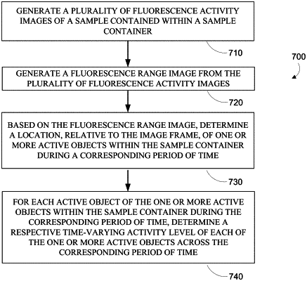
1. A method of imaging and analyzing cell samples in a cell culture vessel, the method comprising:
capturing a movie of the cell culture vessel;
generating a static range image from the movie, wherein the static range image is composed of pixels representing the minimum fluorescence intensity subtracted from the maximum fluorescence intensity at each pixel location over a complete scan period; and
defining objects by segmenting the static range image.