| CPC G01N 21/6458 (2013.01) [G01N 15/14 (2013.01); G01N 21/6402 (2013.01); G01N 21/1702 (2013.01); G01N 33/582 (2013.01); G01N 2800/56 (2013.01); G02B 21/0076 (2013.01)] | 24 Claims |
|
1. A method of detecting ovarian circulating tumor cells in a subject suspected of having cancer, the method comprising:
obtaining a test sample from the subject;
contacting the test sample with nanoparticles, wherein the nanoparticles are folic acid functionalized nanoparticles further comprising a fluorescent label;
incubating the test sample with the nanoparticles for a period of time sufficient to allow uptake of the nanoparticles by the ovarian circulating tumor cells in the test sample;
removing from the test sample any free nanoparticles not taken up by an ovarian circulating tumor cell;
illuminating the test sample with laser light to excite the test sample in the capillary tube; and
detecting the presence of the ovarian circulating tumor cells in the subject's sample when photoacoustic signals generated by the nanoparticles are detected by displaying the photoacoustic signals as a photoacoustic image;
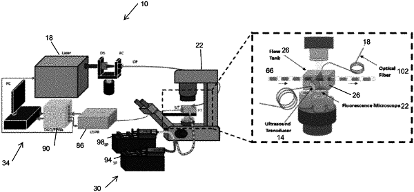
wherein the method is performed using a photoacoustic flow system comprising:
a flow chamber configured to support a capillary tube, the flow chamber including a window and a slot;
a pump system coupled to the capillary tube, the pump system including a first syringe pump filled with air and a second syringe pump containing the test sample, wherein the first pump injects the air into the capillary tube, and the second pump injects the test sample into the capillary tube to produce two-phase flow with alternating air and test sample through the capillary tube;
an optical fiber coupled to a laser and configured to transmit the laser light that excites the nanoparticles uptaken by the ovarian circulating tumor cells in the test sample in the capillary tube;
an ultrasound transducer coupled to the window of the flow chamber, wherein the ultrasound transducer detects the photoacoustic signals generated by the excited nanoparticles;
an inverted fluorescence microscope including a stage for supporting the flow chamber and a camera aligned with the slot of the flow chamber for obtaining images of the test sample as it passes through the capillary tube; and
a data acquisition system configured to receive the photoacoustic signals from the ultrasound transducer and the images from the camera to reconstruct and display the photoacoustic image.
|