| CPC A61B 34/10 (2016.02) [A61B 5/0075 (2013.01); A61B 5/7246 (2013.01); A61B 17/3421 (2013.01); A61B 34/20 (2016.02); A61B 34/25 (2016.02); G06T 7/0014 (2013.01); G06T 7/30 (2017.01); G16H 10/40 (2018.01); G16H 20/40 (2018.01); G16H 30/20 (2018.01); G16H 30/40 (2018.01); G16H 40/20 (2018.01); G16H 50/20 (2018.01); G16H 50/50 (2018.01); G16H 50/70 (2018.01); A61B 90/50 (2016.02); A61B 2034/101 (2016.02); A61B 2034/105 (2016.02); A61B 2034/107 (2016.02); A61B 2034/2051 (2016.02); A61B 2034/2055 (2016.02); A61B 2034/252 (2016.02); A61B 2034/256 (2016.02); A61B 2090/103 (2016.02); A61B 2090/365 (2016.02); A61B 2090/374 (2016.02); A61B 2090/378 (2016.02); A61B 2090/3762 (2016.02); A61B 2090/571 (2016.02); A61B 2576/026 (2013.01); G06T 2207/30016 (2013.01); G16H 10/60 (2018.01); G16H 70/20 (2018.01)] | 20 Claims |
|
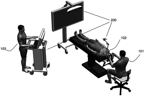
1. A method of performing intraoperative biopsy tracking during at least one medical procedure, by way of an automatic system comprising: a control and processing system comprising a processor, a tracking system, and a tracking engine interfaced with the processor and the tracking system; a storage device interfaced with the control and a processing system; and an external user input and output device interfaced with the control and a processing system comprising providing a display device, the method comprising:
comparing, according to pre-selected similarity criteria, biopsy analysis data and archival biopsy analysis data associated with at least one medical procedure by using the processor, comparing comprising:
detecting and identifying a plurality of local features in images relating to the biopsy analysis data and images relating to the archival biopsy data by using computer vision;
detecting and identifying a plurality of local features in an initial medical image by using a scale-invariant feature transform algorithm to facilitate searching biopsy analysis data and archival biopsy data, using the scale-invariant feature transform algorithm comprising: producing a difference image from the initial medical image by blurring the initial medical image, thereby producing a blurred image; subtracting the blurred image from the initial medical image, thereby producing the difference image; locating pixel amplitude extrema in the difference image; defining a corresponding pixel region about each pixel amplitude extremum of the pixel amplitude extrema; producing a plurality of component subregion descriptors for each subregion of a pixel region about the pixel amplitude extrema in the difference image; correlating the plurality of component subregion descriptors for each subregion of the pixel region about the pixel amplitude extrema in the difference image with a plurality of component subregion descriptors for each subregion of a pixel region about the pixel amplitude extrema in the initial medical image; and indicating detection of each local feature of the plurality of local features if a number of component subregion descriptors of the plurality of component subregion descriptors defines an aggregate correlation exceeding a threshold correlation;
reducing a size of the biopsy analysis data and the archival biopsy data by decomposing images relating to a clinical data set using at least one feature extraction algorithm, the clinical data set comprising data relating to at least one of: imaging contrast dynamics, diffusion information, quantitative T1 and T2, CT contrast flow, and PET tracer dynamics, whereby searching the biopsy analysis data and the archival biopsy data is performed in a dimensional space less than that corresponding to all of the biopsy analysis data and the archival biopsy data; and
correlating apparent diffusion coefficient (ADC) with cellularity to build a mathematical model by using additional clinically relevant criteria comprising at least one of: a tumor size, a tumor location, a tumor histology, and a pathology result, thereby transforming measured ADC values into cellularity measures.
|