| CPC A61B 8/0883 (2013.01) [G06T 7/0012 (2013.01); G06V 10/56 (2022.01); G06V 10/82 (2022.01); G16H 30/20 (2018.01); G16H 50/20 (2018.01); G06T 2207/10132 (2013.01); G06T 2207/30048 (2013.01)] | 11 Claims |
|
1. A computer-implemented method of analysing a structure within a patient's heart performed by a software component executing on at least one processor, comprising the steps of:
receiving from a memory, a patient study comprising a plurality of transthoracic echocardiography and/or transesophageal echocardiography images of the patient's heart obtained by an ultrasound device;
extracting respective sets of pixel data from the transthoracic echocardiography and/or transesophageal echocardiography images;
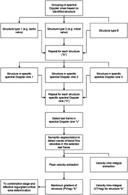
using one or more trained machine learning algorithms to analyse the sets of pixel data to
a) identify first transthoracic echocardiography and/or transesophageal echocardiography images captured using a colour Doppler modality and assign one or more of the first transthoracic echocardiography and/or transesophageal echocardiography images to the structure,
b) identify second transthoracic echocardiography and/or transesophageal echocardiography images captured using spectral Doppler modality and assign one or more of the second transthoracic echocardiography and/or transesophageal echocardiography images to the structure,
c) determine a flow convergence zone radius R from an analysis of the first transthoracic echocardiography and/or transesophageal echocardiography images assigned to the structure; and
d) determine a maximum gradient of structure, PVreg, from an analysis of the second transthoracic echocardiography and/or transesophageal echocardiography images assigned to the structure; and
combining the flow convergence zone radius, R, the maximum gradient of structure PVreg, and a Nyquist value Va identified for the colour Doppler modality, to determine an Effective Regurgitant Orifice Area, EROA for the structure according to the formula:
(2*π*R**2×Va)/PVreg.
|