| CPC A61B 6/504 (2013.01) [A61B 5/0066 (2013.01); A61B 5/0075 (2013.01); A61B 5/055 (2013.01); A61B 5/7267 (2013.01); A61B 5/742 (2013.01); A61B 5/7475 (2013.01); A61B 6/032 (2013.01); A61B 6/037 (2013.01); A61B 6/463 (2013.01); A61B 6/467 (2013.01); A61B 6/481 (2013.01); A61B 6/5205 (2013.01); A61B 8/12 (2013.01); A61B 8/14 (2013.01); A61K 49/04 (2013.01); G06F 18/10 (2023.01); G06T 7/0012 (2013.01); G06V 10/20 (2022.01); G06V 10/245 (2022.01); G06V 10/761 (2022.01); G06V 10/764 (2022.01); G06V 10/82 (2022.01); G06T 2207/10081 (2013.01); G06T 2207/10088 (2013.01); G06T 2207/10101 (2013.01); G06T 2207/10132 (2013.01); G06T 2207/20081 (2013.01); G06T 2207/30048 (2013.01); G06T 2207/30101 (2013.01); G06V 10/247 (2022.01)] | 22 Claims |
|
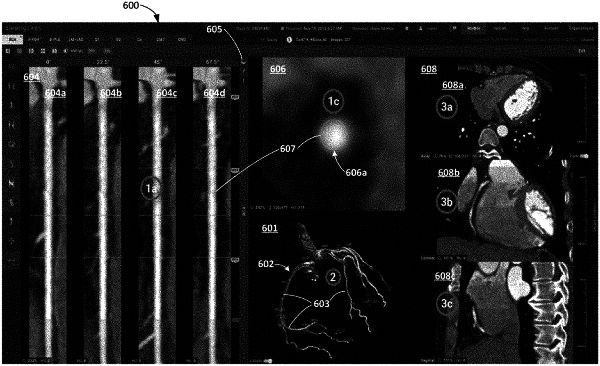
1. A computer-implemented method for displaying computed tomography (CT) images and corresponding coronary vessel information including images rendered from the CT images and identification of lumen, vessel walls, and plaque of coronary vessels determined from the CT images by image processing, the method comprising:
accessing, by a computer system, a set of CT images of coronary vessels of a subject and coronary vessel information associated with the set of CT images, the coronary vessel information including identification of lumen, vessel walls, and plaque of one or more coronary vessels;
generating and displaying, by the computer system, in a user interface, a first panel illustrating at least a portion of a coronary vessel of the subject in a plurality of straightened multiplanar (SMPR) vessel views comprising the coronary vessel information, wherein the coronary vessel information includes a color overlay that depicts calcified plaque with a first color and non-calcified plaque with a second color, the plurality of SMPR vessel views adjacently positioned to one another in the first panel, each of the plurality of SMPR vessel views rotationally offset from one another by a predetermined rotational angle along a longitudinal axis of the plurality of SMPR views;
generating and displaying, by the computer system, in the user interface, a second panel showing a cross-sectional view of the portion of the coronary vessel and the coronary vessel information, wherein the cross-sectional view is generated using the set of CT images, and wherein locations along the plurality of SMPR vessel views are associated with a CT image in the set of CT images such that a selection of a particular location along the portion of the coronary vessel in the plurality of SMPR vessel views displays the associated CT image in the cross-sectional view in the second panel;
receiving, by the computer system, a user input on the user interface indicating a particular location along the portion of the coronary vessel of the subject in the plurality of SMPR vessel views, wherein the user input comprises selecting a particular point or slice along the portion of the coronary vessel in the plurality of SMPR vessel views; and
in response to the user input, displaying, by the computer system, in the second panel, the CT image in the set of CT images that is associated with the location along the portion of the coronary vessel in the cross-sectional view, wherein the associated CT image includes the color overlay depicting calcified plaque with the first color and non-calcified plaque with the second color,
wherein the computer system comprises a computer processor and an electronic storage medium.
|