| CPC G06T 5/009 (2013.01) [G06T 5/40 (2013.01); G06T 11/005 (2013.01); G06T 11/008 (2013.01); G06T 2207/10081 (2013.01); G06T 2207/30008 (2013.01)] | 15 Claims |

|
1. A method for obtaining a computer tomography (CT) image of an object, comprising:
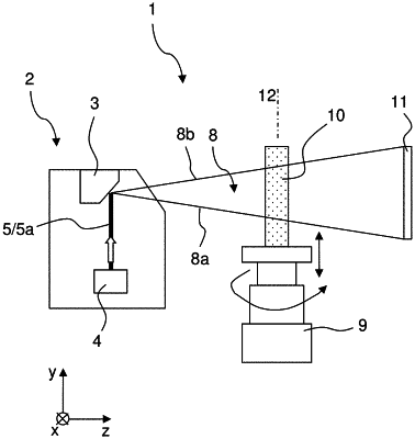
a) generating x-rays using an x-ray source comprising an angled anode, wherein the x-rays are generated within an anode material of the angled anode, and the x-rays exiting the anode material have travelled different distances (Pla, PLb) within the anode material depending on an exit location with respect to a y-direction;
b) recording at least one set of 2D projections of the object or a part of the object located within a beam path of the generated x-rays with a 2D x-ray detector located in the beam path behind the object, with the x-ray source and the 2D x-ray detector being part of a measurement setup, and with the 2D projections of a respective set of 2D projections being recorded one by one in different rotation positions of the object relative to the measurement setup, wherein the different rotation positions of the object relative to the measurement setup are with respect to a turning axis parallel to the y-direction, and wherein for each respective set of 2D projections, a respective shifting position of the object relative to the measurement setup including the x-ray source and the 2D x-ray detector with respect to the y-direction is chosen;
c) generating, for each respective set of 2D projections, at least one 3D CT image of the object or said part of the object, wherein the 3D CT image consists of a plurality of voxels having respective grey values, where the respective grey values represent a respective local x-ray attenuation of the object; and
d) for each generated 3D CT image, correcting the 3D CT image into a corrected 3D CT image or correcting a 2D CT partial image of the 3D CT image into a corrected 2D CT partial image,
wherein for each slice of voxels of the 3D CT image, a scaling factor (sf) is determined, wherein each slice of voxels comprises voxels of the 3D CT image having an identical position in the y-direction, wherein the grey values of each voxel of a respective slice of voxels of the 3D CT image or the 2D CT partial image are multiplied with the scaling factor (sf) determined for the respective slice of voxels, resulting in corrected grey values for the voxels of the corrected 3D CT image or the corrected 2D CT partial image, and
wherein the scaling factors (sf) for the slices of voxels of the 3D CT image are determined with at least one 3D CT calibration image measured with the measurement setup, wherein the at least one 3D CT calibration image depicts similar or identical object structures of a calibration object placed within the beam path of the x-rays in different regions (R1, R2) of a field of view of the measurement setup with respect to the y-direction, wherein for the at least one 3D CT calibration image, a grey value contribution to the grey values of voxels belonging to the similar or identical object structures in said different regions (R1, R2) attributable to a slice position (ns, js) in the y direction is determined, and the scaling factor (sf) for a respective slice of voxels is chosen such that it compensates for the determined grey value contribution for the respective slice of voxels in order to establish a heel effect compensation.
|