| CPC G01R 33/4828 (2013.01) [G01R 33/4838 (2013.01)] | 18 Claims |
|
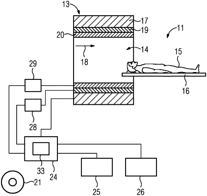
1. A method for actuation of a magnetic resonance (MR) device comprising a radio frequency (RF) antenna configured to generate an RF pulse for specific saturation of nuclear spins in an examination region of an examination object, the method comprising:
generating, via control circuitry, a frequency spectrum of the examination region;
generating, via the control circuitry, a B0 field map;
determining, via the control circuitry, a first resonance frequency for a first tissue and a second resonance frequency for a second tissue based upon the frequency spectrum;
selecting, via the control circuitry, a partial region of the examination region;
determining, via the control circuitry, a saturation pulse using an RF pulse configured for a spectrally-selective excitation of the first tissue and the second tissue based upon the first resonance frequency, the second resonance frequency, and the B0 field map, the determination of the saturation pulse being based upon (i) the spectrally-selective excitation of the first tissue in the examination region, and (ii) the spectrally-selective excitation of the second tissue in the partial region;
generating, via the control circuitry, the saturation pulse via the RF antenna, the saturation pulse comprising part of an MR control sequence;
executing the MR control sequence to generate MR signals;
acquiring, via the MR device, raw data using the MR signals; and
reconstructing, using the acquired raw data, a medical image of the examination region of the examination object.
|