| CPC A61F 2/30756 (2013.01) [A61B 17/1615 (2013.01); A61B 17/1635 (2013.01); A61B 17/1675 (2013.01); A61F 2/30 (2013.01); A61F 2/30771 (2013.01); A61F 2/4618 (2013.01); A61F 2002/305 (2013.01); A61F 2002/3085 (2013.01); A61F 2002/30367 (2013.01); A61F 2002/30387 (2013.01); A61F 2002/30405 (2013.01); A61F 2002/30462 (2013.01); A61F 2002/30485 (2013.01); A61F 2002/30784 (2013.01); A61F 2002/30841 (2013.01)] | 19 Claims |
|
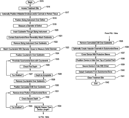
1. A method of preparing a defect at a treatment site on a subchondral bone to repair an anatomical joint and ameliorate a joint condition, the method comprising:
surgically accessing the treatment site;
accessing a sizing instrument, the sizing instrument having a proximal end, a distal end, and a cylindrical member disposed between the proximal end and the distal end, the cylindrical member having a lumen for receiving a guidewire;
centrally positioning the distal end of the sizing instrument over the defect at the treatment site on the subchondral bone; the distal end having concentric rings with each ring having a known diameter;
measuring a diameter of the defect by comparing the defect diameter with the closest corresponding known diameter on the sizing instrument;
inserting the guidewire through the lumen of the cylindrical member while the distal end of the sizing instrument is centrally positioned over the defect;
contacting the subchondral bone with a distal end of the guidewire;
marking the center of the defect by reversibly attaching the guidewire to the subchondral bone;
removing the sizing instrument over the guidewire;
selecting a countersink instrument having a diameter substantially matching the measured diameter of the defect;
positioning the countersink instrument over the guidewire;
simultaneously rotating and lowering the countersink instrument to engage a soft tissue and the subchondral bone;
penetrating the subchondral bone with the countersink instrument to form a hole, the hole having a first depth;
removing the countersink instrument over the guidewire;
positioning a cannulated drill over the guidewire;
removing an inner circular portion of the subchondral bone while preserving a central post of subchondral bone in a substantially undisturbed native state by simultaneously rotating and lowering the cannulated drill into a center of the hole, the inner circular portion having a second depth;
removing the cannulated drill over the guidewire;
placing an implantable orthopedic device in the hole over a top of the central post; wherein the central post is accepted by a hollow central column of the implantable orthopedic device when implanted;
securing the implantable orthopedic device into the hole using a driver instrument;
removing the driver instrument;
detaching the guidewire from the subchondral bone; and removing the guidewire.
|