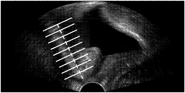
| CPC A61B 8/085 (2013.01) [A61B 5/7267 (2013.01); A61B 8/463 (2013.01); A61B 8/467 (2013.01)] | 18 Claims |

|
1. A method for measuring a parameter in an ultrasound image, comprising:
obtaining a pelvic ultrasound image with an ultrasound probe, wherein the pelvic ultrasound image contains an area representing the pelvic floor tissue;
displaying, by an image processor, the pelvic ultrasound image on a display device;
automatically determining, by the image processor by using a pattern recognition model, a position of an inferoposterior margin of symphysis pubis in the pelvic ultrasound image;
automatically determining, by the image processor, a horizontal axis according to the position of the inferoposterior margin of symphysis pubis;
automatically determining, by the image processor by using the pattern recognition model, a position of a bladder neck in the pelvic ultrasound image, wherein the pattern recognition model is obtained by training one of a ascade adaBoost detector using Haar features, a cascade adaBoost detector using Local Binary Patterns features, a support Vector Machine detector, or a detector based on neural network with positive image samples containing the inferoposterior margin of symphysis pubis and the bladder neck and negative image samples not containing the inferoposterior margin of symphysis pubis and the bladder neck; and
calculating, by the image processor, a distance from the position of the bladder neck to the horizontal axis to obtain a value of a bladder neck-symphyseal distance.
|