| CPC A61B 3/14 (2013.01) [G02B 21/0032 (2013.01); G02B 21/365 (2013.01); G06T 7/0012 (2013.01); G06T 2207/30041 (2013.01)] | 6 Claims |
|
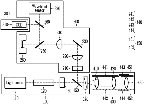
1. A retina imaging device comprising:
a light irradiation unit for irradiating two dispersed lights;
a magnification adjustment unit for adjusting the paths of the two lights and adjusting a magnification of an image obtained from the two lights incident on the eyeball, the magnification adjustment unit comprising:
a first lens on which the two dispersed lights are incident
a second lens on which the two lights that have passed through the first lens are incident; and
a third lens on which the two light that have passed through the second lens are incident, wherein the magnification of the image is defined according to a movement of the third lens such that a first movement of the third lens provides a first magnification and a second movement of the third lens provides a second magnification;
a light compensation unit for receiving lights reflected from the eyeball and compensating for aberrations of lights generated in the eyeball; and
a light processing unit comprising a charge-coupled device (CCD) that converts the compensated reflected lights into an electrical signal, the light processing unit configured for obtaining differential interference contrast (DIC) images by converting the electrical signal of the compensated reflected lights into a digital signal and image-processing the digital signal,
wherein the retina imaging device is configured to obtain at least one eyeground image at the first magnification, and obtain a plurality of DIC images at the second magnification higher than the first magnification with respect to the retina of the entirety of the obtained at least one eyeground image.
|