| CPC A61B 1/2736 (2013.01) [A61B 1/000094 (2022.02); A61B 1/000095 (2022.02); A61B 1/00186 (2013.01); G06T 5/40 (2013.01); G06T 7/0012 (2013.01); G06T 2207/10024 (2013.01); G06T 2207/10068 (2013.01); G06T 2207/20024 (2013.01); G06T 2207/30092 (2013.01)] | 17 Claims |
|
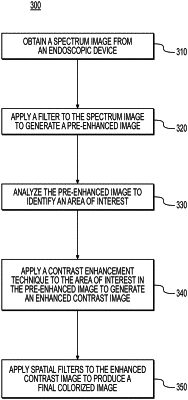
1. A method for enhancing medical images, the method comprising:
at a processor device in communication with an endoscopic device, obtaining from the endoscopic device a spectrum image of bleeding in an upper gastrointestinal (GI) area of a patient;
at the processor device, applying a filter to the spectrum image to generate a pre-enhanced image, wherein the filter enhances the spectrum image at one or more light wavelengths in the light spectrum, and the pre-enhanced image includes three spectral filters at 490 nm, 630 nm, and 640 nm;
analyzing the pre-enhanced image to identify an area of interest, wherein the area of interest represents a portion of the upper GI area with an active bleed;
applying a contrast enhancement technique to the area of interest in the pre-enhanced image to generate an enhanced contrast image; and
applying spatial filters to the enhanced contrast image to produce a final colorized image with defined blood vessels in the upper GI area of the patient.
|